
1. Introduction
The most appropriate process to prevent the spread of bacteria, fungi, and viruses is to wash your hands using soap and running water. Get used to using masks or washing hands with soap or antiseptic hand sanitizer gel preparations (hand sanitizer) after every activity [1, 2]. Antiseptic is a hand sanitizer product with the main content of alcohol, which can kill or inhibit the growth of microorganisms [3]. Excessive use of alcohol can result in increased skin permeability by eliminating lipids in the stratum corneum layer, which can trigger Systematic Contact Dermatitis (SCD) [4].
The use of antiseptic raw materials in general is still dominated by using alcohol, so alternative raw materials are needed that can be used as antiseptics, especially hand sanitizers other than alcohol. One of the materials under consideration is liquid smoke. In general, liquid smoke is the result of condensation or condensation of vapour from biomass combustion through a pyrolysis process. This combustion is carried out
either indirectly or directly from materials that contain a lot of carbon and other compounds [5]. The selection of liquid smoke as an antiseptic raw material comes from previously conducted research. It is known that liquid smoke from bamboo stems [6], coffee skin [7], bamboo leaves [8], pine fruit [9], and palm oil [10] can be an antiseptic.
The effectiveness of liquid smoke as an antiseptic is because liquid smoke contains antibacterial compounds, namely phenols and acids. These phenol and acid fractions can inhibit the growth of microorganisms [11]. Considering some research results that show that the chemical content, especially phenol, in liquid smoke functions as a disinfectant, it is possible to conduct further research on the utilization of tealeaf liquid smoke as an antiseptic. Tea leaf (Camellia sinensis) is one of the natural ingredients that can be used as an antiseptic because it has phenol compounds that can dam- age mycobacterial cell membranes [12]. Therefore, this study aims to determine the effectiveness of tea-leaf liquid smoke as an antiseptic raw material.
2. Methodology
2.1. Preparation of Tea Leaf Liquid Smoke
The tea leaves utilized as the primary ingredient for liquid smoke are the trimmings discarded from five-year-old tea plants sourced from the Indonesia Research Institute for Tea and Cinchona. The raw material in the form of dried tea leaves is weighed as much as 3 kg, then put into a pyrolysis furnace made of stainless steel equipped with a cylindrical pyrolysis tube (retort) with a height of about 60 cm and a diameter of 40 cm equipped with 2 thermocouples, an electric heating device, a condenser, and a distillate collection flask. Pyrolysis was carried out at 250–450 °C for 7-8 hours. The pyrolysis process is carried out in a special furnace that does not allow the involvement of oxygen in the pyrolysis process [13]. The initial pyrolysis liquid smoke (grade 3) still contains tar, acidity, and odor. Because the smoke is highly unpleasant, it must be filtered using the distillation procedure to generate grade 1 and grade 2 liquid smoke. Grade 2 liquid smoke is obtained by distilling grade 3 liquid smoke once, while grade 1 liquid smoke is obtained by distilling grade 3 liquid smoke twice [10].
2.2. In Vitro Testing
Escherichia coli ATCC 25922, Staphylococcus aureus ATCC 25923, Candida albicans ATCC 10231, and Aspergillus flavus ATCC 9643 were investigated in vitro for their antibacterial effectiveness. The test microbes used were identified by gram staining for bacteria and methyl blue staining for fungi. Microbial identification employs colony morphology, cell morphology, and gram staining techniques to verify the absence of contamination in the utilized microbes. To determine the ability of tea leaf liquid smoke to inhibit bacteria using the disc paper method while inhibiting fungi using the well method with a 6 mm diameter disc paper [14]. The work was done under sterile conditions to avoid contamination from other microbes.
The inhibition test used grade 1 and 2 tea leaf liquid smoke with concentrations of 35%, 50%, and 75%. Observations on the media were made after 24 hours of incubation. The diameter of the inhibition zone, or clear zone, that appears around the disc paper is an indication of the microbial sensitivity to the antimicrobial material used as test material and is expressed by the diameter of the inhibition zone. The zone of inhibition formed around the disc paper was measured by vertical diameter and horizontal diameter in mm units using a caliper [15].
Table 1. Microbial inhibition response based on clear zone diameter [16]
| Diameter of clear zone (mm) | Microbial inhibition response | |
|---|---|---|
| < 5 | Weaker | |
| 5 - 10 | Medium | |
| 10 - 20 | Strong | |
| > 20 | Very strong | |
2.3. In Vivo Testing
In vivo tests were carried out by administering liquid smoke to the palms of respondents according to the grade and optimal concentration of the in vitro test results [8]. The number of research samples was 24 respondents, who weredivided into two groups of 12 people each for the administration of liquid smoke as a test material and 70% alcohol as a control. Swabbing of respondents' palms was carried out before and after antiseptic application, and then the results of the swab suspension were planted on nutrient agar (NA) medium and potato dextrose agar (PDA) medium in a pour plate to calculate the effectiveness of microbial reduction. The questionnaire parameters were liking for color, aroma, dryness effect, itching effect, and burning [10].
2.4. Data Analysis
Microsoft Excel organizes and evaluates data in a table format, including measurements of the inhibition zone, calculations of colony numbers, and questionnaire evaluations of tea-leaf liquid smoke as an antiseptic.
3. Result and Discussion
3.1. Physical Properties of Liquid Smoke
The yield of liquid smoke produced from the pyrolysis process of tea leaves using a reactor with a capacity of 3 kg is 46.28%. The pyrolysis apparatus, the length of the combustion, the kind of raw material, and the initial moisturecontent of the raw tea leaf materials all have an impact on the quality and yield of liquid smoke. The liquid smoke used in this study is distilled liquid smoke at a temperature of 100–190°C. This distillation aimsto separate the carcinogenic tar con- tent in the liquid smoke. The physical properties of tea-leaf liquid smoke can be seen in Table 2, which include color, aroma, and pH.
Table 2. Physical properties of liquid smoke
| Physical properties | Liquid smoke type | |||
|---|---|---|---|---|
| properties | Grade 3 | Grade 2 | Grade 1 | |
| Color | Brownish-yellow | Clear yellow | Translucent yellow | |
| Aroma | Strong smoke odor | Smells lighter of smoke | Smells lighter of smoke | |
| pН | 5,4 | 2,8 | 2,7 | |
The distilled liquid smoke (grade 2 and grade 1) has a lighter color than the liquid smoke before distillation (grade 3). This happens because grade 3 liquid smoke still contains impurities such as tar, while grade 2 and 1 liquid smoke have reduced tar and benzopyrene content. The aroma produced in liquid smoke before and after distillation remains the same, which has a distinctive odor [17]. The purification of liquid smoke to produce grade 2 liquid smoke is done by distillation at a temperature of 130–145 °C for 6 hours. At this stage,distillation is carried out to increase the acquisition of acetic acid and ensure that there are no more impurities, such as tar and benzopyrene. The liquid smoke produced in this grade is clear yellow, but the odor of liquid smoke in grade 2 is lighter than in grade 3. The pH value in grade 2 also increased to 2.8 due to the increase in acid content in grade 2 liquid smoke.
Grade 1 liquid smoke is obtained by distilling the pyrolyzed liquid smoke at 170-190°C for 4 hours. The purpose of this stage of distillation is to increase the recovery of acidand phenol content in liquid smoke and ensure that there are no impurity compounds in this grade so that it is safe to use(18). The grade 1 liquid smoke produced has a clearer color than the other grades and has a lighter smoke aroma as well when compared to the smoke from the other grades'liquids. The pH value of this grade is 2.7, indicating that the acid content and phenol content of the liquid smoke are increasing.
3.2. Identification of Test Microbes
Microbial identification was carried out on Escherichia coli ATCC 25922, Staphylococcus aureus ATCC 25923, Candida albicans ATCC 10231, and Aspergillus flavusATCC 9643 macroscopically and microscopically. The results of macroscopic and microscopic observations can be seen in Table 3 below.
Table 3. Results of macroscopic and microscopic observations of test microbes
| No | Microbe type | Morphology of colonies | Cell morphology | Staining result |
|---|---|---|---|---|
| 1. | Escherichia coli ATCC 25922 | Round, smooth, convex, milky white, flat | Stem, monobacilli | Colored red |
| 2. | Staphylococcus aureus ATCC 25923 | Round, smooth, convex, milky white, flat | Rod, monobacillus | Purple-colored |
| 3. | Candida albicans ATCC 10231 | Round, white in color, convex, soft, flat | ound, purple, has pseudohyphae and blastophores | Purple-colored |
| 4. | Aspergillus flavus ATCC 9643 | Round filamentous, white (young) and greenish (old), convex | Spherical with short conidia stalks | Blue colored |
Based on the results in Table 3, it appears that E. coli ATCC 25922 is a gram-negative bacterium because it has a thin cell wall that is between two layers of the cell membrane. S. aureus is a gram-positive bacterium because it has a thick cell wall, a cell membrane layer, and no outer membrane [19]. Gram staining results show that S. aureus ATCC 25923 is purple because the crystal violet dye is retained in alcohol, as opposed to E. coli ATCC 25922, which is red because the crystal violet dye fades in alcohol and allows the safranin dye to be absorbed [20].
The results of macroscopic observations of C. albicans ATCC 10231 have round colonies, white in color with smooth textured colonies, and convex elevations, thisis because C. albicans is a yeast [21]. The results of gram staining of C. albicans ATCC 10231 with oval-shaped, purple-colored colonies indicate a gram-positive bacterial group. These gram-positive
bacteria are able to retain crystal violet dye. The macroscopic observation of A. flavus ATCC 9643 is a round-shaped colony, with a colony size of 0.5 - 2 cm, with a white color when incubated for 24 hours and a greenish color when incubated for 48 hours, convex elevation and has a velvety texture. Meanwhile, microscopic results with methyl blue staining, the results obtained round spores with short conidia stalks. Methyl blue staining in fungi will distinguish which yeast cells are still alive and which are dead because methyl blue will give col- or when oxidation occurs. This reduction is what causes the color to disappear and oxidation will cause a blue color [22].
The data obtained from microbial identification, whichincludes the examination of colony morphology, cell morphology, and gram staining, confirms that all microbial cultures are free from impurities and contaminants. This ensures that the selected treatment will produce optimal outcomes for the specific microorganism being targeted.
3.3. In Vitro Test
The presence of a clean zone around the disk or well indicates the outcome of the antimicrobial activity test. The results of in vitro testing of tea-leaf liquid smoke can be seen in Table 4. Based on Table 4, it can be seen that the diameter of the clear zone formed is getting bigger as the concentration increases. This means that the greater the concentration, the greater the diameter of the clear zone produced. Lala [23] stated that the higher the concentration of liquid smokeadded to the product, the lower the acid value, or pH. The higherthe concentration of ingredients, the higher the content of antibacterial active substances. The addition of antibacterial compound concentrations is thought to increase the penetration of antibacterial compounds into microbial cells, which will damage the cell's metabolic system and can result in cell death.
Table 4. Results of measuring the diameter of the liquid smoke inhibition zone against test microbes
| Liquid smoke | Concentration (%) | Test microbial species and inhibition zone diameter (mm) | |||
|---|---|---|---|---|---|
| type | Escherichia coli | Staphylococcus | Candida | Aspergillus | |
| ATCC 25922 | aureus ATCC | albicans | flavus ATCC | ||
| (mm) | 25923 | ATCC 10231 | 9643 | ||
| (mm) | (mm) | (mm) | |||
| Grade 1 | 35 | 1,3 (L) | 2,3 (L) | 0 (L) | 9,8 (S) |
| 50 | 1,8 (L) | 3,0 (L) | 1,0 (L) | 16,1 (K) | |
| 75 | 3,0 (L) | 5,8 (S) | 1,5 (L) | 17 (K) | |
| Grade 2 | 35 | 1,3 (L) | 4,8 (L) | 0,8 (L) | 16,1 (K) |
| 50 | 2,0 (L) | 5,8 (S) | 1,5 (L) | 23,3 (SK) | |
| 75 | 3,5 (L) | 9,0 (S) | 3,8 (L) | 25,6 (SK) | |
Based on the inhibition response criteria according to Andriani et al. [16], the grade 1 and 2 tea leaf liquid smoke against Escherichia coli ATCC 25922 has an inhibition response that is classified as weak (<5 mm), against Staphylococcus aureus ATCC 25923 classified as weak to moderate (5- 10 mm), against Candida albicans ATCC 10231 classified as weak, and against Aspergillus flavus ATCC 9643classified as moderate to very strong (>20 mm).
Liquid smoke has inhibition against microbial growth, which can be caused by the content of phenol and acetic acid, which can function as antibacterial. The mechanism of phenol compounds and their derivatives as disinfectants is to damage bacterial cell membranes causing leakage of organic ions, nucleotides, coenzymes, and amino acids out of bacterial cells and prevent essential substances from entering the cell. This causes disruption of the bacterial growth system and can cause cell death. Acetic acid is one of the organic acids commonly used as an antibacterial because it is able to reduce pH, causing instability in the cell membrane [17].
Phenol compounds and their derivatives easily form protein complexes through hydrogen bonds. At low levels, protein-phenol complexes will form weak bonds and immediately decompose into cells, so that protein denaturation occurs in the bacterial cell wall. At high levels, the presence of phenolic compounds can cause bacterial cells to lyse [24]. Acetic acid in liquid smoke also has an important role as an antibacterial because it has an effect on destabilizing the function and structure of components in bacterial cells [25].
3.4. In Vivo Test
.
The test of the effectiveness of tea-leaf liquid smoke as an antiseptic in vivo was carried out by applying tea-leaf liquid smoke to the palms of the respondents. This in vivo test was conducted to support the results of the liquid smoke effectiveness test on microbial growth in vitro. The research respondents were 24 people consisting of 2 groups, namely respondent group A (swab the respondent's palm before and after using 70% alcohol) as many as 12 respondents and respondent group B (swab the respondent's palm before and after using 75% tea leaf liquid smoke) as many as 12 respondents, in each group evenly distributed male and female respondents.
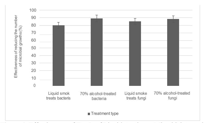
Figure 1. Effectiveness of Tea Leaf Liquid Smoke on Microbial Growth
In order to calculate the percentage of the efficacy of tea leaf liquid smoke in preventing bacterial and fungal growth, the number of bacterial and fungal colonies decreased during the in vivo effectiveness test of tea leaf liquid smoke as an antiseptic. The results in Figure 1 show that the 75% tea leaf liquid smoke treatment decreased the number ofbacterial colonies by 80% and decreased the number of fungalcolonies by 85%, while the 70% alcohol treatment showed a decrease in the number of bacterial colonies by 89% and fungi by 87.2%.
The decrease in the amount of microbial growth with tealeaf liquid smoke indicates that tea-leaf liquid smoke has the ability to inhibit microbial growth. The results of thisstudy are in accordance with research conducted by Oktarina et al. [26] that showed that liquid smoke containing phenol compounds and acetic acid effectively inhibits the growth of Escherichia coli. Compound phenol in liquid smoke can form phenol-protein complex bonds that cause protein coagulation so that the cell membrane is lysed. According to Erlytasari et al. [27], phenol compounds contained in liquid smoke can bind to bacterial proteins through hydrogen bonds, causing the protein structure to be damaged.
The decrease in the number of fungal colonies using 75% tea-leaf liquid smoke was lower compared to 70% alcohol. This shows that 70% alcohol is still superior in inhibiting the growth of fungal colonies compared to 75% tea-leaf liquid smoke. According to research conducted by Putri et al. [28], antiseptics containing 70% alcohol content will be more sensitive and work faster to precipitate proteins and lipid membranes in microbes. In addition, 70% alcohol is considered superior to inhibiting microbial growth because 70% alcohol is obtained from the results of purification through several stages, including evaporation, distillation, dehydration, and recryption stages known as the refinery process (29).
3.5. Questionnaire Results
In this study, questionnaire data was also collected from respondents regarding their level of preference for the physical characteristics of tea-leaf liquid smoke. This evaluation was performed to see if the direct application of tea-leaf liquid smoke to the respondent's hand had any effects. In Figure 2, it can be seen that overall respondents liked the color of tea leaf liquid smoke (92%), liked the aroma of tea leaf liquid smoke (75%), liked the tea leaf liquid smoke product because it did not give the effect of dryness on the hands (92%), and liked the bamboo stem liquid smoke product because it did not give the effect of a burning sensation (100%).
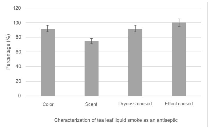
Figure 2. Questionnaire for Respondents' Assessment of Tea Leaf Liquid Smoke
Based on Figure 2, 92% of respondents liked the color of the 75% concentration of tea leaf liquid smoke with yellowish brown color criteria. Aznuri et al. [30] stated thatthe level of preference for the color of the antiseptic gel is green and bright because it more appealing compared to dark colors that are less attractive to the public. In this study, tea-leaf liquid smoke added 1% orange perfume and increased the respondents' level of preference for the antiseptic aroma of tea-leaf liquid smoke by 75%. This is in accordance with the research of Rindarwati and Noviyanto [31]. Regarding consumer interest in the use of antiseptics, people like antiseptics that are attractive and refreshing. This is also similar to research conducted by Handayani et al. [32] regarding the characteristics of antiseptic ingredients that people like, namely that people prefer antiseptics that can provide an attractive aroma, such as the scent of lemon or others. Rindarwati and Noviyanto [31] also stated that the comfort factor of using antiseptic products must prioritize color, texture, and aroma and not cause side effects on the skin.
4. Conclusion
The tea leaf liquid smoke exhibited the most significant inhibition zone when grade 2 liquid smoke with a concentration of 75% was used. Liquid smoke from the pyrolysis of tea leaves (Camellia sinensis) as an antiseptic raw material is effectively used to inhibit the growth of bacteria and fungi.
Acknowledgement
The authors are grateful for the support of the Institut Teknologi Bandung Indonesia for funding this research through the Research and Community Service and Innovation Program (P3MI-2020).