

ISOLASI DAN AKTIVITAS ANTIBAKTERI DARI JAMUR ASAL TANAH TAMAN BOTANI NEGARA, SHAH ALAM, MALAYSIA
ABSTRAK
Jamur merupakan organisme eukariotik yang terdiri dari organisme uniseluler yaitu kapang dan ragi, dan organisme multiseluler yang dikenal sebagai jamur. Jamur memiliki kontribusi yang besar terhadap bidang kesehatan karena merupakan sumber yang banyak digunakan dalam pencarian kandidat antibiotik baru. Tujuan dari penelitian ini adalah mengisolasi jamur yang berasal dari tanah hutan Malaysia sebagai sumber pencarian antibiotik baru dan menentukan aktivitas antibakterinya terhadap Bacillus subtilis dan Escherichia coli. Isolasi jamur dari sampel menggunakan media agar Sabouraud dextrose (SD). Skrining aktivitas antibakteri dilakukan terhadap isolat jamur murni dengan metode difusi agar. Isolat jamur yang aktif selanjutnya di fermentasi dengan medium cair SD selama 21 hari. Kultur media dan miselia dipisahkan dengan menggunakan metode filtrasi. Bagian kultur media di ekstraksi dengan ekstraksi cair-cair (ECC) sedangkan bagian miselium diekstraksi dengan cara maserasi menggunakan pelarut etil asetat. Ekstrak kering diuji aktivitas antimikrobanya terhadap bakteri uji dengan metode mikrodilusi. Hasil dari penelitian ini diperoleh lima strain jamur yang diberi kode S1-S5. Hasil uji aktivitas antibakteri menunjukkan bahwa ekstrak kultur media jamur S3 mempunyai aktivitas antibakteri yang paling tinggi terhadap Bacillus subtilis dengan konsentrasi hambat minimum (KHM) sebesar 64 µg/mL dan jamur S1 terhadap Escherichia coli dengan KHM sebesar of 32 µg/mL. Berdasarkan nilai KHMnya kedua jamur tersebut diklasifikan mempunyai aktivitas antibakteri yang signifikan. Ekstrak jamur tersebut berpotensi berguna untuk pengembangan senyawa terapetik yang baru untuk melawan infeksi bakteri.
Kata kunci: aktivitas antibakteri, Bacillus subtilis, Escherichia coli, isolasi, jamur
Introduction
Majority of fungi produces secondary metabolites which may be beneficial towards pharmaceutical chemist as these metabolites are widely used in medicine such as the development of antibiotics, anti-fungal, and anticholesterol (Zeilinger et al. 2015). For instance, the first well-known antibiotic produced, penicillin, was obtained from the fungi of Penicillium notatum. This discovery was made by Sir Alexander Fleming in 1928. There are hundreds of thousand species of fungi that have been identified. Also, more than millions may exist (Madigan et al. 2012).
One of the prolific sources of fungi is soil. Majority of forest soil community is dominated by fungi. On account of organic compounds in forest soil, fungi species can extend their filaments known as hyphae. A network of hyphae, known as mycelium, is used to absorb nutrients from the ground. Plenteous soil-derived fungi contain hyphae for growth.
This paper focuses on isolation of the soil-derived fungi from Malaysian forest as resources for new antibiotics. Moreover, the investigation of their antibacterial activity against Bacillus subtilis and Escherichia coli through determination the minimum inhibitory concentration (MIC) is presented in this paper.
Materials and Methods
Sample Collection and Preparation
The forest soil was collected in an opened area in Taman Botani Negara, Shah Alam, the state capital of Negeri Selangor Darul Ehsan, Malaysia. The coordinates, latitude, and longitude, of soil collection, are 3° 5'46.20"N and 101°30'42.73"E respectively. The collected soil was placed in a plastic container and kept in a freezer until the start of experimentation.
Isolation and Purification of Soil Derived Fungi
An amount of 1 gram soil sample was weighed and transferred into a centrifuge tube containing 10 mL of sterile water. The mixture was mixed for 1 minute. Then, 1 mL of the mixture was taken and transferred into a test tube containing 9 mL of sterile water. The mixture was gently shaken until become homogenous. From this mixture, 1 mL was taken and transferred into a new test tube containing 9 mL of water. The mixture was stirred gently until homogenous. Then, 1 mL of this mixture was transferred into a sterile petri dish. Lastly, 25 mL of sterile SDA was added into the petri dish and was shaken gently to ensure homogeneity. Once the medium solidified, the petri dish was labeled and incubated at 20⁰C. Fungi grew after 3 to 4 days. The growing fungal was carefully transferred to a new media and repeated until getting a pure culture. Each pure fungal strain was observed under a microscope.
Screening of Antibacterial activity of Pure Fungal Isolates against Test Bacteria
Each pure fungal strains were cut to a size of 1 cm × 1 cm. These fungal strains were then placed on a surface of MHA containing 100 µL bacterial suspensions and incubated at 37 ⁰C for 24 hours. Each fungal strain was observed its capability to inhibit the bacterium growth by measuring the zone of inhibition produced.
Fermentation of Fungal Strains
Each pure fungal isolates were cut to a size of 1 cm × 1 cm and were transferred to a 250 mL Erlenmeyer flask. Sterile SDB with the volume of 100 mL was added into each Erlenmeyer flask to undergo a fermentation process. The mouth of Erlenmeyer flask was covered with cotton which was wrapped with a sterile gauze pad. Fermentation was carried out at room temperature and was placed on a shaker for 21 days.
Extraction
After 21 days of fermentation, the medium was filtered under vacuum using Buchner funnel to separate the mycelium residue and the media culture. The mycelium residue was macerate for 48 hours in 100 mL of EA. On the other hand, the medium culture was extracted using liquid-liquid extraction (LLE) for 3 times using 300 mL of ethyl acetate. The solvent was removed under vacuum (150 mbar) by using a rotary evaporator until an oily residue was formed. The residue was transferred into an empty vial that was weighed. The extract was then dried at room temperature for 24 hours. After a day, the weight of the dried extract formed was measured.
Minimum Inhibitory Concentration (MIC) Assay
The Minimum Inhibitory Concentration was determined using microdilution method (CLSI 2010). The dried extract was dissolve using methanol to achieve a stock concentration of 2048 µg/mL. Each well of the microplates was filled with 100 µL MHB. Extract with the volume of 100 µL and concentration of 2048 µg/mL was added in well number 12. A series of dilution was done by pipetting 100 µL of the mixture from well number 12 to well number 3. From well number 3, 100 µL of mixtures was discarded. Then, the bacterial suspension was added from well number 2 to well number 12. From this series of dilution, well
number 1 and 2 shows negative and positive control respectively. Also, the highest concentration of extract was 1024 µg/mL, located on well number 12, while the lowest concentration of extract was 2 µg/mL, found on well number 3. The microplate was incubated at 37 ⁰C for 24 hours. The minimum inhibitory concentration was measured by observing the turbidity of the mixture after incubation.
Results and Discussion
The morphological characteristic of each pure fungal strain was observed and identified. Some features such as the color, surface, form, elevation, and margin were observed and recorded. The result of morphological characterization was shown in Table 1. Microscopic analysis was done to further analyze the morphological characteristics of each fungal colony, such as the
| Table 1. Morphological Characteristic of Pure Fungal Strain (Leung and Liu 2005). | |||
|---|---|---|---|
| Fungal Isolate | Colour | Surface | Form | Elevation | Margin |
|---|---|---|---|---|---|
| SA 1 | Yellowish green with white edges | Cotton-like | Irregular | Convex | Filiform |
| SA 2 | Light green | Cotton-like | Irregular | Raised | Filiform |
| SA 3 | Greenish blue with white edges | Sand-like | Circular | Convex | Entire |
| SA 4 | White | Smooth | Circular | Raised | Entire |
| SA 5 | Black | Rough | Irregular | Raised | Undulate |
types of spores, hyphae, and mycelium. The results of fungi hyphae observation showed that pure fungal colonies of SA 4 and SA 5 contained septate hyphae, whereas SA 2 contained aseptate hyphae. Different results on the fungal colonies of SA 1 and SA 3, it could not be observed any hyphae on them. The results of spore observation showed that the spores of pure fungal colonies SA 1 and SA 3 were blastospore, whereas pure colonies of SA 2, SA 4, and SA 5 were conidia as illustrated in Figure 2. Therefore, based on the morphological characteristic, the suggested genus of SA 1, SA2, and SA 5 were Aspergillus. On the other hand, the proposed genus of SA 3 and SA 4 were Penicillium and Rhizopus respectively as tabulated in Table 2. In this case, the molecular biological identification is necessary to confirm their species.
Each fungal isolates was tested its capability to inhibit the growth of bacterium surrounding the fungus. Two types of bacteria, gram positive and gram negative, used in this analysis were B. subtilis and E. coli, respectively. The fungi that exhibited a diameter of zone inhibition, namely S1, S3, and S4, (as shown in Figure 3, Table 3) proceeded towards fermentation process to produce their secondary metabolites.
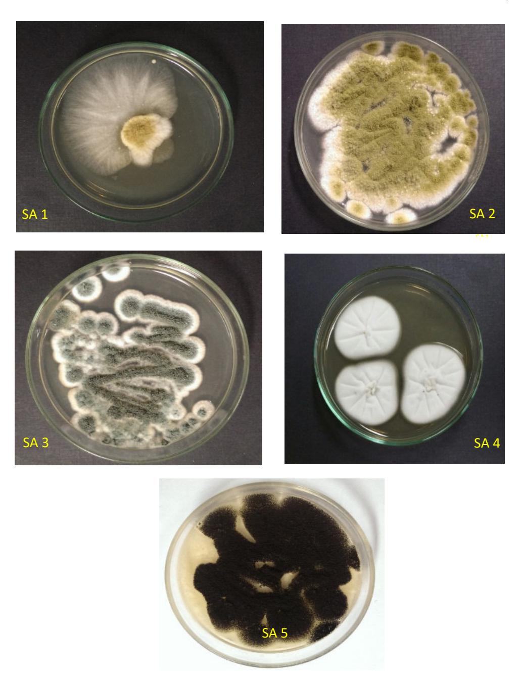
Figure 1. Pure Isolated Colonies of Fungi.
Figure 2. The Microscopic Observation of Pure Fungal Isolates, Magnification 100x.
Table 2. The Microscopic Characteristic of Pure Fungal Strain (Leung and Liu 2005).
| Fungal Isolate | Spore | Hyphae and mycelium |
|---|---|---|
| SA 1 | Blastospore | - |
| SA 2 | Conidium | Aseptate |
| SA 3 | Blastospore | - |
| SA 4 | Conidium | Septum |
| SA 5 | Conidium | Septum |
Figure 3. Zone of Inhibition Produced by Pure Fungal Colony Against B. subtilis (a), E. coli (b).
Table 3. Inhibition Zone Produced by The Pure Fungal Isolates Against Test Bacteria.
| Test bacteria | Inhibition Zone (mm) | ||||
|---|---|---|---|---|---|
| SA1 | SA2 | SA3 | SA4 | SA 5 | |
| Bacillus subtilis | 15.7 | - | 6.6 | 14.9 | - |
| Escherichia coli | 23.1 | - | - | 18.7 | - |
Note : - , no inhibition zone
Extraction of fermentation product was done on both mycelium and liquid medium because fungal secondary metabolites can exist either in extracellular (liquid medium) or intracellular (mycelium residue). Then, the dried extracts of SA 1, SA 3, and SA 4 were used for determining their minimum inhibitory concentration (MIC).
The medium extract of SA 3 gave the lowest MIC value against B. subtilis at 64 µg/mL, followed by SA 1 with MIC 256 µg/mL. However, fungal
medium extract of SA 4 showed growth until the concentration of extract was 2048 µg/mL against B. subtilis and E. coli. The fungal extract medium of SA 1 also gave the lowest MIC against E. coli at 32 µg/mL. However, the fungal medium extract of SA 3 and SA 4 did not show any inhibition against E. coli.
The extract mycelium of S3 and S4 have the same MIC value against B. subtilis at 256 µg/mL, while the SA 1 gave the MIC value of 512 µg/mL. The lowest MIC value against E. coli was shown by extract mycelium of SA 4 at 64 µg/mL, followed by SA 1 with the concentration of 256 µg/mL (Table 4). Meanwhile, fungal extract SA 3 did not give any MIC value against Escherichia coli. The classification of antimicrobial activity of extracts based on MIC value as follows: significant if MIC values are below 100 μg/ml, moderate when 100<MIC<625 μg/mL and weak if MIC>625 μg/mL (Kuete et al. 2011, Dzoyem et al. 2013). From the result shown that fungal
S1 against E. coli. Based on their MIC values, they can be classified as the extracts with significant antibacterial activities.
Table 4. The MIC Value of Fungal Media And Mycelium Extract.
| Fungi | Concentration of extract (µg/mL) | |||||
|---|---|---|---|---|---|---|
| Media Culture | Mycelium | |||||
| B. subtilis | E. coli | B. subtilis | E. coli | |||
| SA 1 | 256 | 32 | 512 | 256 | ||
| SA 3 | 64 | - | 256 | - | ||
| SA 4 | - | - | 256 | 64 | ||
Note: -, MIC > 2048 µg/mL
Among the test bacteria, Bacilus subtilis is the most sensitive to the extracts. Meanwhile, E. coli was the most resistant. The different response towards antibacterial substances is due to their outer membrane. The bacterial envelope of B. subtilis, a Gram positive bacterium, consists of lipoteichoic acid, teichoic acid, peptidoglycan, protein, and phospholipid. The peptidoglycan layer is located on the outermost part or the surface of the bacterium. On the other hand, E. coli, a Gram negative bacterium, consist of lipopolysaccharide, lipoprotein, peptidoglycan, protein, and phospholipid. The structures of gram negative bacterium differ from a gram positive bacterium regarding the location of peptidoglycan. The peptidoglycan in gram negative bacterium is situated in between the outer membrane, which is the cell wall, and the cell membrane (Madigan et al. 2012). This outer layer prohibits certain molecules such as drugs, from penetrating the bacterium cell, consequently making the bacterium more resistant to drugs in contrast with a gram positive bacterium (Kaplan 2000).
Conclusion
A total of five types of soil-derived fungi (SA1- SA5) were isolated from Taman Botani Negara, Shah Alam, Malaysia. Based on the morphological characteristic, the suggested genus of SA 1, SA2, and SA 5 were Aspergillus, whereas SA 3 and SA 4 were Penicillium and Rhizopus, respectively. The antibacterial assay
medium extract of S3 had the highest antibacterial activity against B. subtilis and showed that fungal medium extract of S3 had the highest antibacterial activity against B. subtilis and the extract mycelium of S4 against E. coli. Also based on their MIC values, they were classified as extracts with significant antibacterial activities. These extracts could be potentially useful for the development of new therapeutic agents against bacterial infections.
Acknowledgment
The authors also thank School of Pharmacy ITB-Bandung Institute of Technology for providing the facilities and reagents to conduct this research.
References
Clinical and Laboratory Standard Institute (CLSI), 2010, Method for Dilution Antimicrobial Susceptibility Test for Bacteria that Grow Anaerobically, Approved Standard M07-A8 8th ed.; National Committee for Clinical Laboratory Standards: Wayne, Philadelphia, USA.
Dzoyem JP, Guru SK, Pieme CA, Kuete V, Sharma A, Khan IA, Saxena AK, Vishwakarma RA, 2013, Cytotoxic and antimicrobial activity of selected Cameroonian edible plants, BMC Complementary and Alternative Medicine, 13 (78): 1-6.
Kaplan M, 2000, Melissa Kaplan's Herp care collection: the problem with gram-negative bacteria, http://www.anapsid.org/gramnegative.html, accessed on 3rd May 2016.
Kuete V, Krusche B, Youns M, Voukeng I, Fankam AG, Tankeo S, Lacmata S, Efferth T, 2011, Cytotoxicity of some Cameroonian spices and selected medicinal plant extracts, J Ethnopharmacol 134:803–812.
Madigan M, Martinko J, Stahl D, Clark D, 2012, Brock biology of microorganisms, 13th edn, Pearson, San Fransisco.
Zeilinger S, Martin J, Estrada C, 2015, Biosynthesis and molecular genetics of fungal secondary Metabolites, 2nd edn, Springer, New York.