

STUDI NMR METIL β‐ORSELLINAT DARI DIRINARIA SP. YANG DIAMBIL DARI KAWASAN PANTAI TELUK NIPAH, PULAU PANGKOR, PERAK MALAYSIA.
ABSTRAK
Penelitian dilakukan untuk mengisolasi dan mengkarakterisasi metil β‐orsellinate dari lichen Dirinaria sp. yang diambil dari daerah pesisir Teluk Nipah, Pulau Pangkor, Perak Malaysia. Sampel diekstraksi dalam metanol sebanyak tiga kali pada suhu ruang. Selanjutnya, ekstrak kasar dianalisis menggunakan kromatograϐi lapis tipis (KLT) dan kromatograϐi cair vakum (KCV). Untuk mendapatkan senyawa murni dilakukan tahap isolasi dengan menggunakan kromatograϐi radial (KR). Struktur kimia senyawa isolat ditentukan dengan menggunakan beberapa spektroskopi yaitu infra merah (IM), resonansi magentik inti (NMR), dan spektrometri massa (MS). Senyawa tersebut diidentiϐikasi sebagai metil β‐orsellinate.
Kata kunci: metil β‐orselinat, liken, Dirinaria sp.
INTRODUCTION
Lichen is a form organism of a symbiotic relationship between fungal and a photosynthetic organism like blue‐green algae or cyanobacteria (Bebert 1831). Lichens could grow almost anywhere, although the type of species may differ based on environmental conditions (Nimis et al. 2002; Nash 2008; Norela et al. 2018). Pulau Pangkor, Perak, is a tropical island with near‐ constant exposure to the sun and wind. This research was done at Teluk Nipah, lat. 4°N, long. 100°E. The types of lichens mainly grow in this environment must be hardy, resistant to moisture loss, have ultraviolet‐light tolerant and salt‐tolerant properties (Kosanićet al. 2010). Dirinaria sp. which from the family of Physciaceae was reported that it has produced many types of aromatic compounds ( Samsudin 2010; Kekuda et al. 2015; Abas et al. 2019). The present study was performed to report the isolation and characterization of methyl β‐ orsellinate using 1D and 2D NMR spectroscopy.
MATERIALS AND METHODS
General
Thin‐layer chromatography, aluminium sheets 20 × 20 cm of the silica gel 60 F254 of 0.25 mm thickness (art. no. 5554, MERCK) and Silica gel; Kieselgel 60 PF254 (art. no. 7749, Merck) (radial chromatography, Chromatotron‐7924T‐01 USA). The structure of the isolated compound was determined based on the spectral data recorded on FTIR (Perkin‐Elmer USA) spectrophotometer and NMR 400 MHz (JEOL). ESI‐MS was recorded by using Ultra performance liquid chromatography (model JEOL JMS‐700/GI.)
Lichen Material
The sample used in this research was the lichen Dirinaria sp. which was collected from coastal areas of Teluk Nipah, Pulau Pangkor, Perak Malaysia.
Extraction and Isolation
The ground air‐dried of Dirinaria sp. (700 g) was
Figure 1. TLC Proϐile of Dirinaria sp. Spot with the box is the isolated compound
extracted for 250 ml X 3 days with ethyl acetate at room temperature.. The EtOAc extract was concentrated at 40‐50 °C with a rotary evaporator under reduced pressure to give 150 g green pale extract. The crude extract was fractionated by using vacuum liquid chromatography (VLC) eluted with increasing polarity of n‐hexane‐EtOAc. The eluents that showed the same proϐile on thin layer chromatography (TLC) chromatogram were combined to give three fractions (I‐III). Puriϐication of Fraction III (300mg) was carried out using Radial Chromatography (RC) with silica gel plate of 1 mm thickness eluted with 95:5 n‐hexane‐EtOAc in 5% polarity increment to yield compounds 1 (30 mg) (Rosandy et al. 2013). All the prior proϐile wasdetected using smaller pieces of TLC and then detected under UV light (254 nm) (A) and by H2SO4 spraying reagent followed by heating (B) (Figure 1).
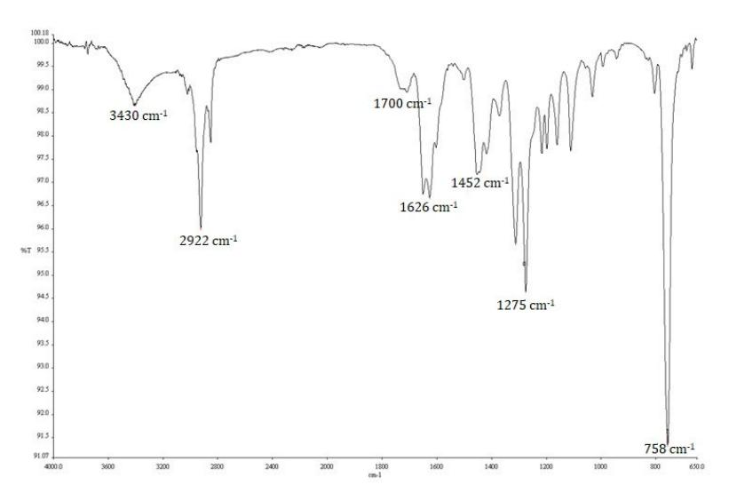
Figure 2. IR spectrum of compound 1.
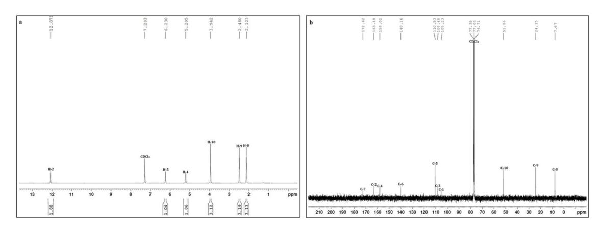
Figure 3. 1H(a) and 13C(b)‐NMR spectrum of compound 1.
Methyl β‐orsellinate (1) MP: 142‐144 °C. EI‐MS [M]+ at m/z 196.077 IR (MeOH) νmax (cm‐1): 3430, 2922, 1700, 1626, 1275, and 758. 1H NMR (Chloroform‐d, 844 MHz): Table 1 13C NMR (Chloroform‐d, 100 MHz): Table 1
RESULTS AND DISCUSSIONS
Puriϐication with vacuum liquid chromatography (VLC) was isolated compound 1 as a yellow solid, molecular ion peak EI‐MS [M]+ at m/z 196.07 and melting point at 142‐144 °C. The IR spectrum (Figure 2.) displayed absorption bands at 3430 cm‐1 (OH group) and 2922 cm‐1 (sp3 C‐H group), 1700 cm‐1 (C=O group), 1626 cm‐1 (C=C aromatic), 1275 cm‐1 (C‐O ether) and 758 cm‐1 (‐CH3‐ group bonding).
The 1H NMR (Figure 3.) for compound 1 showed three signals for methyl group at δH 2.12 ppm (3H, s, H‐8) and at δH 2.48 ppm (3H, s, H‐9), a methoxy group at δH 3.94 ppm (3H, s, 10‐OCH3), an aromatic proton at δH 6.23 ppm (1H, s, H‐5), and two
hydroxyl protons at δH 12.07 ppm (1H, s, 2‐OH) and δH 5.21 ppm (1H, s, 4‐OH). The 13C NMR spectrum (Figure 2.) showed 10 signals corresponding to two methyl group at δc 7.7 ppm (C‐8) and at δC 24.2 ppm (C‐9), a methoxy group at δC 51.9 ppm (10‐ OCH3), six aromatic carbons at δC 105.2 ppm (C‐1), δC 163.2 ppm (C‐2), δC 108.5 ppm (C‐3), δC 158.0 ppm (C‐4), δC 110.5 ppm (C‐5), δC 140.2 ppm (C‐6), an ester group at δC 172.6 ppm (C‐7). Based on this data, the structure of 1 has a molecular formula of C10H12O4 and DBE (Double Bond Equivalent) value is 5, indicating that the compound consists of four double bonds (three oleϐinic and one carbonyl) and one ring. The complete assignment of protonated carbon was conϐirmed by HSQC correlations (Table 1.;Figure 3.). It was evident from the correlation there were two methyls, one methoxy and a methine proton in the molecule.
The HMBC spectrum of 1 (Table 1; Figure 4b.) showed long‐range correlations between aromatic proton at δH 6.23 (H‐5) with aromatic carbons at δC 105.2 (C‐1), 108.5 (C‐3), 158.0 (C‐4) and methyl proton δC 24.2 (C‐9), the hydroxy proton at δH 12.07 (2‐OH) with aromatic carbon at δC 105.2 (C‐
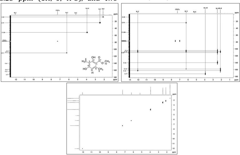
Figure 4. 1Hಫ 13C HSQC (a), 1H↔13C HMBC (b), and 1H↔1H COSY (c) NMR spectrum of compound 1.
Table 1. 1D and 2D NMR of Compound 1
| No | 13C (ppm) | 13C* (ppm) | 1H↔13C HSQC (∑H, mult. J in Hz) | 1H↔13C HMBC | 1H↔1H COSY | |
|---|---|---|---|---|---|---|
| 1 | 105.2 | 105.2 | ‐ | ‐ | ‐ | |
| 2 | 163.2 | 163.1 | ‐ | ‐ | ‐ | |
| 2‐OH | ‐ | ‐ | 12.07 (1H, s) | C‐1, C‐2, C‐3, C‐4 | ‐ | |
| 3 | 108.5 | 108.5 | ‐ | ‐ | ‐ | |
| 4 | 158.0 | 158.0 | ‐ | ‐ | ‐ | |
| 4‐OH | ‐ | ‐ | 5.21 (1H, s) | ‐ | ‐ | |
| 5 | 110.5 | 110.5 | 6.23(1H, s) | C‐1, C‐3, C‐4, C‐9 | ‐ | |
| 6 | 140.2 | 140.2 | ‐ | ‐ | ‐ | |
| 7 | 172.6 | 172.6 | ‐ | ‐ | ‐ | |
| 8 | 7.7 | 7.6 | 2.12 (3H, s) | C‐2, C‐3, C‐4 | ‐ | |
| 9 | 24.2 | 24.1 | 2.48 (3H, s) | C‐1, C‐5, C‐6 | ‐ | |
| 10‐OCH3 | 51.9 | 51.8 | 3.94(3H, s) | C‐7 | ‐ |
1), 163.2 (C‐2), 108.5 (C‐3) and 158.0 (C‐4) indicating that all methyl and hydroxy groups were attached to the aromatic ring. The long rage correlation of proton methyl at δH 3.94 (H‐10) with carbonyl carbon at δC 172.6 (C‐7) suggested the attachment of this carbonyl to the aromatic ring to form methyl benzoate. Based on the facts above, the structure of 1 is determined as methyl 2,4‐ dihydroxy‐3,6‐dimethylbenzoate or methyl β‐ orsellinate (Hylands and Ingolfsdottir 1985; Toledo et al. 6447; Huynh et al. 6459; Perico‐Franco et al. 2015).
CONCLUSION
A methyl β‐orsellinate was successfully isolated from ethyl acetate extract lichen Dirinaria sp was collected from coastal areas of Teluk Nipah, Pulau Pangkor, Perak Malaysia. The chemical structure of the isolated compound was elucidated by using IR, 1D and 2D NMR spectroscopy and MS spectrometric technique
ACKNOWLEDGEMENT
We would like to thank the School of Chemical Sciences and Food Technology, Faculty of Science and Technology, Universiti Kebangsaan Malaysia (UKM) for providing research facilities and express our gratitude to the Ministry of Higher Education Malaysia and UKM for ϐinancial support through research grants of FRGS/1/2014/SG01 UKM/2/06.
REFERENCES
Bebert, 1831, Sur une nouvelle substance decouverte dans Ie lichen vulpinus. Rapporteurs: Robiquet et Blondeau, J Pharm Sci Access 17: 696‐ 700.
Nimis PL, Scheidegger C, Wolseley, PA, 2002, Monitoring with Lichens, Springer, Netherland.
Nash TH, 2008, Lichen Biology, 4th Edition, Cambridge University Press, Cambridge.
Norela S, Faezah SFMF, Nasution, SAAM, Nusaibah S, Ismail BS, 2018, Use of lichens as bioindicators for determining atmospheric heavy metal concentration in Malaysia, PakJ Bot 50(1): 421‐428. Kosanić M, Ranković B, Sukdolak S, 2010, Antimicrobial activity of the lichen Lecanora frustulosa and Parmeliopsis hyperopta and their divaricatic acid and zeorin constituents, Afr J Microbiol Res 4: 885–890.
Samsudin MW, 2010, Liken: Tumbuhan primitif yang masih relevan, Universiti Kebangsaan Malaysia, UKM Press, Malaysia.
Kekuda T, Dhanya R, Dhatri R, Sunita C, Onkarappa R, Vinayaka K, 2015, Radical scavenging, antimicrobial and insecticidal efϐicacy of Parmotrema cristiferum and Dirinaria applanate, Science, Technology and Arts Research Journal, 4 (1): 95.
Abas A, Khalid RM, Rosandy AR, Norela S, 2019, Lichens of pulau Pangkor, Perak, Malaysia, The
Malaysian Forester 82 (1): 59‐66.
Rosandy AR, Din LB, Yaacob WA, Yusoff NI, Sahidin I, Latip J, Nataqain S, Noor NM, 2013, Isolation and characterization of compounds from the stem bark of Uvaria rufa (annonaceae), The Malaysian Journal of Analytical Sciences 17(1): 50‐58.
Hylands PJ, Ingolfsdottir K, 1985, The isolation of methyl β‐Orsellinate from Stereocaulon alpinum and comments on the isolation of 4,6‐dihydroxy‐2‐ methoxy‐3‐methylacetophenone from Stereocaulon Species, Phytochemistry 68 (5): 56¹ – 29.
Toledo FJM, Garcıá AC, Estévez FR, Quintana JA, Bermejo JB, 2003, Identiϐication and quantitation of allelochemicals from the lichen Lethariella canariensis: phytotoxicity and antioxidative activity, Journal of Chemical Ecology 29(9): 2049‐2071.
Huynh BLC, Le DH, Takenaka Y, Tanahashi T, Nguyen KPP, 2016, New phenolic compounds from the lichen Parmotrema praesorediosum (Nyl.) Hale (Parmeliaceae), Magn Reson Chem 54: 81–87.
Perico‐Franco LS, Soriano‐Garcıá M, Cerbón MA, González‐Sánchez I, Valencia‐Islas NA, 2015, Secondary metabolites and cytotoxic potential of Lobariella pallida and Stereocaulon strictum var. compressum, two lichens from Colombian páramo region, UK Journal of Pharmaceutical and Biosciences 3(4): 31‐38.