

*Corresponding author
Muhamad Insanu E-mail: insanu@fa.itb.ac.id
Active chemical compounds from medicinal plants are secondary metabolites that can be histochemically tested. It enables the identification and localization of diverse secondary metabolites within the tissue, which is not possible with screening phytochemicals. In this study, the histochemistry of the leaf blades of Ericales plants i.e., cantigi (Vaccinium varingiaefolium (Blume) Miq.), persimmon (Diospyros kaki Thunb.), sapodilla (Manilkara zapota (L.) van Royen), and star apple (Chrysophyllum cainito L.) were evaluated. These species have been reported as folk medicines that have secondary metabolites. Each leaf tissue was observed under a microscope, and phytochemical compounds' presence and location were detected using specific reagents. The results showed that those four leaves contained phenolic compounds, flavonoids, and terpenes, while quinones were only found in cantigi leaves. The secondary metabolites were present in the idioblast of parenchymal and epidermal tissues. The persimmon and star apple leaves had secretory cavities in trichomes with terpenes and phenolic compounds.
Key Words: Ericales, histochemistry, secondary metabolites.
ANALISIS HISTOKIMIA EMPAT DAUN TUMBUHAN OBAT DARI ORDO ERICALES
ABSTRAK
Senyawa kimia aktif tanaman obat sebagian besar merupakan metabolit sekunder yang dapat diuji secara histokimia. Metode ini memungkinkan identifikasi dan lokalisasi golongan senyawa metabolit sekunder di dalam jaringan, yang tidak mungkin dilakukan dengan skrining fitokimia. Pada penelitian ini dilakukan analisis histokimia pada daun tumbuhan bangsa Ericales yaitu cantigi (Vaccinium varingiaefolium (Blume) Miq.), kesemek (Diospyros kaki Thunb.), sawo (Manilkara zapota (L.) Van Royen), dan sawo duren (Chrysophyllum cainito L.). Spesies tumbuhan ini telah dilaporkan sebagai obat tradisional yang memiliki metabolit sekunder. Jaringan daun dapat diamati di bawah mikroskop dan keberadaan serta lokasi senyawa metabolit sekunder dideteksi dengan menggunakan pereaksi spesifik. Hasil penelitian menunjukkan bahwa keempat daun tersebut mengandung fenol, flavonoid, dan terpen, sedangkan kuinon hanya terdapat pada daun cantigi. Metabolit sekunder ditemukan terutama pada idioblas sel parenkim dan epidermis. Daun kesemek dan daun sawo duren memiliki sel sekresi pada trikoma dengan kandungan terpen dan fenol.
Kata kunci: Ericales, histokimia, metabolit sekunder
INTRODUCTION
Active chemical compounds are generally included in secondary metabolites such as alkaloids, flavonoids, and terpenes (Dewick 2009). Most of these compounds are stored in secretory structures, while only a limited amount of secondary metabolites are stored in vacuoles and the cytosol of parenchymal cells (Dickison 2000). One way to determine the distribution of secondary metabolites in plants is through histochemical tests, which allow quick and inexpensive preliminary tests in searching for active chemical compounds (Coelho et al. 2012, Royo et al. 2015).
Histochemistry has a tremendous long-term impact on cell and tissue biology, embryology, and pathology and continues at the forefront of research in these disciplines. Histochemical techniques are now more widely used than ever and, in conjunction with new developments in microscopical imaging and analysis, will continue to have an important position in the life sciences and medicine (Coleman et al. 2000). The histochemical analysis is related to the localization and identification of compounds, metabolic activity, and aspects of cell biology from cells and tissues. This technique is used to visualize the type of compounds present in tissues (Lavis 2011). The histochemical test's advantage is locating where the metabolites are produced or accumulated in plant tissues (Mulyani and Laksana 2011).
Among the Angiosperms plant group, Ericales is one of the plant's orders used in traditional medicine for various diseases. The main chemical compounds are triterpenoids and flavonoids (Rocha et al. 2015). According to APG IV system (2016), Ericales comprise about 22 families, Balsaminaceae, Marcgraviaceae, Tetrameristaceae, Fouquieriaceae, Polemoniaceae, Lecythidaceae, Sladeniaceae, Pentaphylacaceae, Sapotaceae, Ebenaceae, Primulaceae, Theaceae, Symplocaceae, Diapensiaceae, Styracaceae, Sarraceniaceae, Roridulaceae, Actinidiaceae, Clethraceae, Cyrillaceae, Ericaceae, and Mitrastemonaceae. Several common medicinal plants of Ericales found in Indonesia are cantigi (Vaccinium varingiaefolium (Blume) Miq.), persimmons (Diospyros kaki Thunb.), sapodilla (Manilkara zapota (L.) van
Royen.), and star apple (Chrysophyllum cainito L.). The leaves of V. varingiaefolium (Ericaceae) are traditionally used for the treatment of wrinkles, external wounds, burns, and inflammation (Yulyana et al. 2016). D. kaki(Ebenaceae) is used as an antioxidant, anti-inflammatory, hypolipidemic, antidiabetic, and antibacterial (Xie et al. 2015). The leaves of M. zapota (Sapotaceae) are useful for the treatment of inflammation, skin diseases, diarrhea, and improving blood circulation (Ganguly and Rahman 2013). Leaf infusion of C. cainito (Sapotaceae) is used to treat diabetes and articular rheumatism (Shailajan and Gurjar 2014).
To our knowledge, the pharmacognostic analysis including phytochemical and histochemical studies on Ericales is still limited. Therefore, this study aimed to analyze the accumulated metabolite compounds in the secretory structure and the phytochemical content in four medicinal plants of Ericales using histochemical tests.
MATERIALS AND METHODS
Leaves of V. varingiaefolium (Blume) Miq., D. kaki Thunb., M. zapota (L.) Van Royen, and C. cainito L. were collected in Bandung. Specimen was deposited at the Herbarium Bandungense, School of Life Sciences and Technology Institut Teknologi Bandung.
The transverse section for histochemical experiments were sectioned by hand-sliced from fresh leaves (3rd node) with three replications. The sample was prepared using a polystyrene mount according to the method of the American Herbal Pharmacopeia (2011). The sample is carefully sliced by running the blade evenly along the flat top surface of the mount during the sectioning technique, which uses a very sharp razor blade. The fresh section is stained in a few drop of reagent solution for 2Ȃ5 minutes, carefully washed with water, and covered with a cover slip. The stained sample was observed under the microscope with magnification of 100Ȃ400x. The specimen measurements are taken by calibrating an eyepiece with an integrated eyepiece micrometer with a precisely measured stage micrometer.
Histochemical tests were performed using the following reagents: 5% ferric chloride (Johansen 1940) and 10% potassium dichromate (Gabe 1968) for phenolic compounds; aluminum chloride (Guerin et al. 1971), citroboric acid (Pedro et al. 1990), sodium hydroxide (Robinson 1983), and vanillin-HCl (Mace and Howell, 1974) for flavonoids; cupric acetate (Rupa 2015) for terpenes; potassium hydroxide (Farnsworth 1966) for quinones; Dragendorff and Wagner (Furr and Mahlberg 1981) for alkaloids. The fresh and unstained sections were used as a negative control. The positive control was performed according to the reference of the respective author. The presence of brown or dark brown color in the tissue indicated a positive test for phenolic compounds. The positive result for flavonoids was indicated by the appearance of yellow with aluminum chloride, citroboric acid, sodium hydroxide, and pink with vanillin-HCl. A positive test for terpenoids was shown by the appearance of yellow or brownish-yellow color in the tissue. The presence of red color in the tissue indicated a positive test for quinone compounds. Alkaloid presence was shown by the presence of reddishbrown or yellow deposits. Image recording was performed using a light microscope (Olympus CX21LED) coupled with a digital camera (SXY-I50L). The histochemical test slides were then analyzed in the Laboratory of Pharmacognosy at the Institut Teknologi Bandung (ITB) for a detailed
description and secondary metabolite of the leaf tissues
RESULTS AND DISCUSSION
Upon histochemical analysis on the transverse section of leaves from four Ericales plants, we found the presence of flavonoid, terpenes, and phenol containing tissues/ cells, and the absence of alkaloids (Table 1). In case alkaloid is present, the cells would be stained reddish-brown with Wagner's reagent ȋMercadante-Simoes et al. 2014, Barbosa et al. 2021) and stained brown with Dragendorff (Matias et al. 2016, Gomez et al. 2019), which did not show in this present study. Meanwhile, quinones were only detected in V. varingiaefolium leaves. Secondary metabolites can be stored in internal and external secretory structures (Dickison 2000). In this study, the internal secretory structure was found in the midrib (the central vein of a leaf) and intercostal (the part between the leaf bone and leaf edge) area, especially in idioblast cells. The idioblast cells that contain the metabolites are in the epidermal tissue, parenchyma, and vascular tissue of all four leaves. The external secretory structures such as trichomes, especially glandular trichomes, are only found on D. kaki, and C. cainito leaves.
Table 1. Results of histochemical tests for the secretory structures on the leaf blade of the four species studied
| Sec'da"› me–ab'li–e• | Reage–• | Sec"e–'"› •–"—c–—"e• | S'ecie• | |||
|---|---|---|---|---|---|---|
| VǤ ˜a"ingiaefoli—m | DǤ kaki | MǤ œa'o–a | CǤ caini–o | |||
| Phenolic compounds | Ferric chloride | Idioblast | Ϊ | Ϊ | Ϊ | Ϊ |
| Potassium dichromate | Idioblast | Ϊ | Ϊ | Ϊ | Ϊ | |
| Flavonoids | Aluminum chloride | Idioblast | Ϊ | Ϊ | Ϊ | Ϊ |
| Citroboric acid | Idioblast | Ϊ | Ϊ | Ϊ | Ϊ | |
| Sodium hydroxide | Idioblast | Ϊ | Ϊ | Ϊ | Ϊ | |
| Vanillin-HCl | Idioblast | Ϊ | - | Ϊ | - | |
| Terpenes | Cupric acetate | Trichomes | - | Ϊ | - | Ϊ |
| Quinones | Potassium hydroxide | Idioblast | Ϊ | - | - | - |
| Alkaloids | Dragendorff | - | - | - | - | - |
| Wagner | - | - | - | - | - | |
Note: (+) = positive result; (-) = negative result
The transverse sections of V. varingiaefolium leaves without reagent solution is showed in Figure 1(A-D). The chemical compounds were visualized by adding various reagent solutions as in Figure 1(E-N). Phenolic compounds were observed in idioblast cells, especially in the midrib and intercostal areas (Figure 1E-F). Flavonoid compounds in idioblast cells are found in the parenchyma of the intercostal (Figure 1H-J) and midrib area (Figure 1K). Quinone compounds showed positive results in palisade and spongy parenchyma of the intercostal site (Figure 1G). The terpenoid compounds were detected in the lower epidermis (Figure 1L). No histochemical were found in the literature to demonstrate the chemical nature of V. varingiaefolium. The chemical compound of V. varingiaefolium leaves has been reported by Yulyana et al. (2016) with phytochemical screening. The positive result showed flavonoid, saponin, steroid/triterpenoid, and tannin. Anthraquinone was not present based on the phytochemical screening test. The difference in the results of this quinone compound was due to the growing location of V. varingiaefolium in Yulyana's research, which was obtained from East Java, Indonesia. In contrast, this study was obtained from West Java, Indonesia. Plants that grow in different places will synthesize other secondary metabolites depending on many factors, such as light, temperature, soil water, soil fertility, salinity, the presence, or absence of stress, etc (Yang et al. 2018).
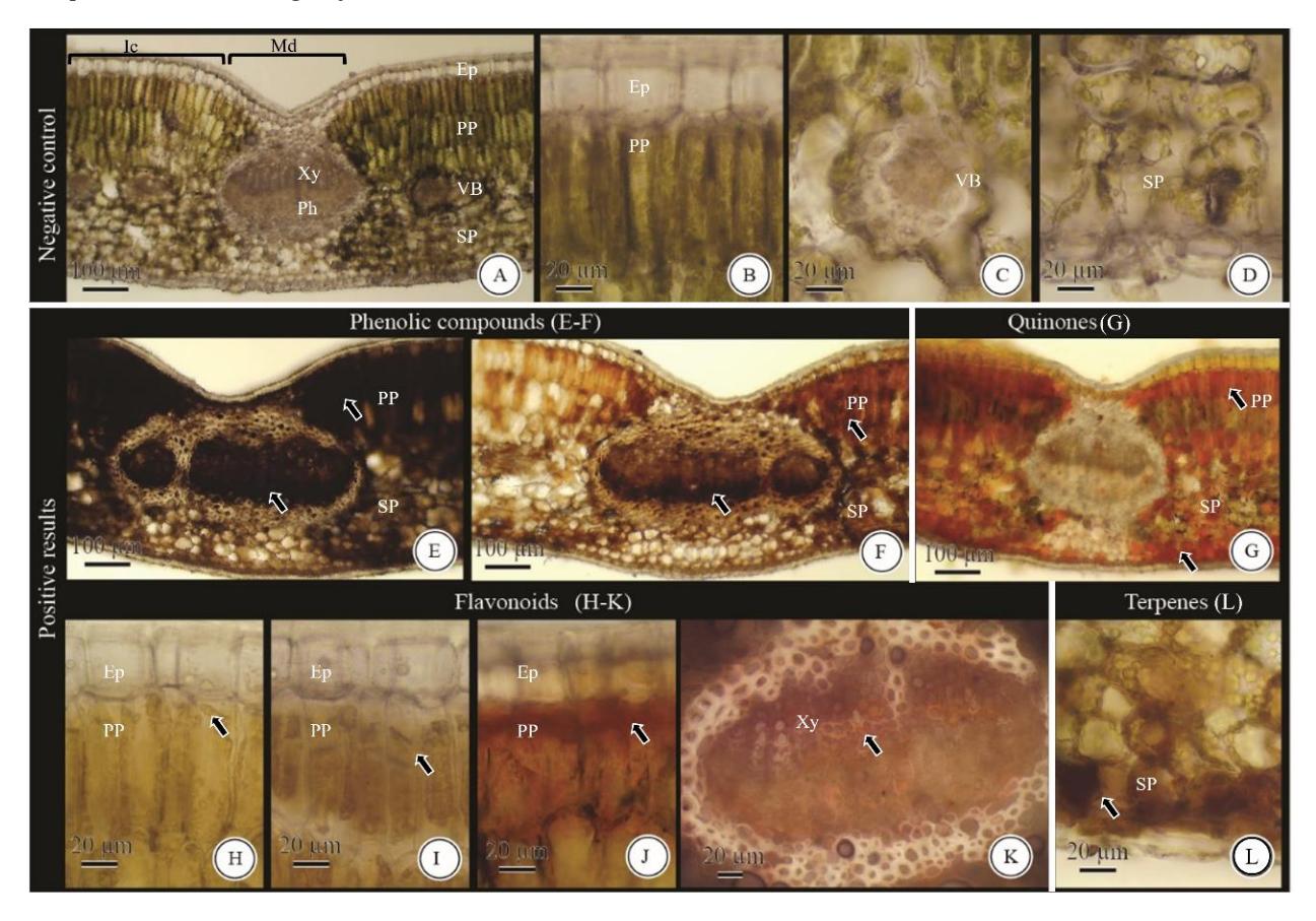
Figure 1. Histochemical characteristic of V. varingiaefolium. (A-D) Cross-section as a negative control. (E-F) Phenolic compounds (E) dark brown with ferric chloride (F) brown with potassium dichromate. (G) Quinones stained red with potassium hydroxide. (H-K) Flavonoids stained (H-J) yellow with (H) aluminum chloride (I) citroboric acid (J) sodium hydroxide (K) pink with vanillin-HCl. (L) Terpenes stained brown with cupric acetate. Ep, epidermis; Ic, intercostal; Md, midrib; Ph, phloem; PP, palisade parenchyma; SP, spongy parenchyma; VB, vascular bundle; Xy, xylem. Arrows indicate idioblasts with chemical compounds.
In D. kaki leaves, the unstained section as the negative control is shown in Figure 2A-E. The histochemical tests showed phenolic compounds in the midrib and intercostal area (Figure 2F; 2I) and the external secretory structure as glandular trichomes (Figure 2G-H; 2J-K). Flavonoid compounds were in idioblast cells of the epidermis and parenchyma in the intercostal
(Figure 2L-N) and midrib areas (Figure 2O). The terpenoid compounds were detected in the glandular trichomes (Figure 2P-Q). These histochemical results align with the previous results based on phytochemical screening conducted by Zreen et al. (2019). Both tests showed tannins (phenolic compounds), flavonoids, and terpenoids.
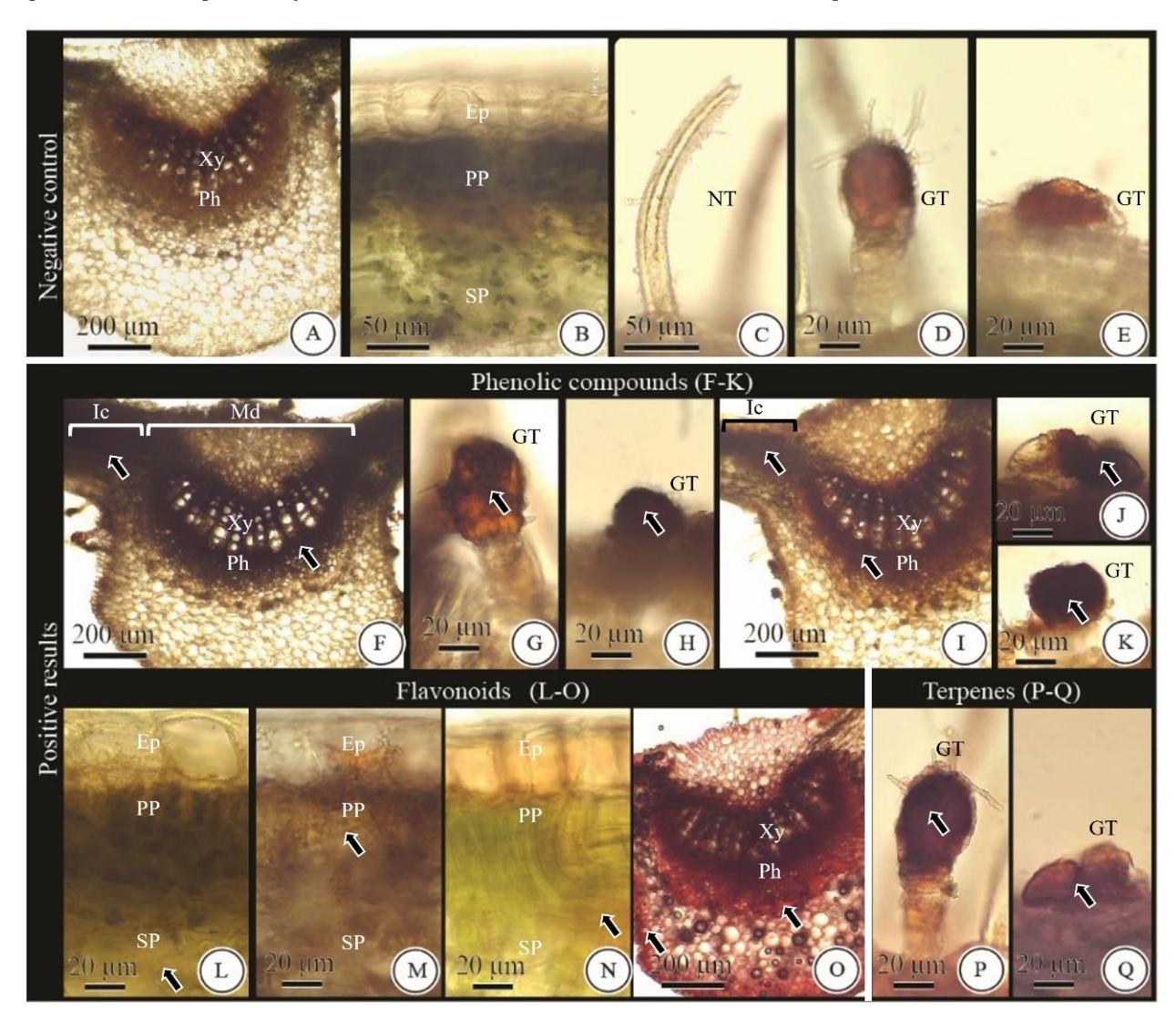
Figure 2. Histochemical characteristic of D. kaki (A-E) Cross-section as negative control. (F-K) Phenolic compounds (F-H) dark brown with ferric chloride (I-K) brown with potassium dichromate. (L-O) Flavonoids stained (L-N) yellow with (L) aluminum chloride (M) citroboric acid (N) sodium hydroxide (O) pink with vanillin-HCl. (P-Q) Terpenes stained brown with cupric acetate. Ep, epidermis; GT, glandular trichome; Ic, intercostal; Md, midrib; NT, non-glandular trichome; Ph, phloem; PP, palisade parenchyma; SP, spongy parenchyma; Xy, xylem. Arrows indicate idioblasts with chemical compounds.
C. cainito presented transverse sections of leaves before adding the reagent (Figure 3A-E). Phenolic compounds were detected in the intercostal area
(Figure 3H-I) and the external secretory structure as non-glandular trichomes (Figure 3F-G; 3J-K). Flavonoid was observed in idioblast cells of the epidermis, parenchyma in the intercostal (Fig. 3L-N), and midrib area (Figure 3O). The terpenoid compounds were in the parenchyma and spongy parenchyma of the intercostal area (Figure 3P). Phytochemical screening (Zuhro et al. (2016) exhibited saponin, triterpenoid, flavonoid, and phenolic compounds in C. cainito leaves. It is similar with our findings which showed that through histochemical examination, C. cainito leaves contained flavonoids, phenolic compounds, terpenoids and not contained alkaloids.
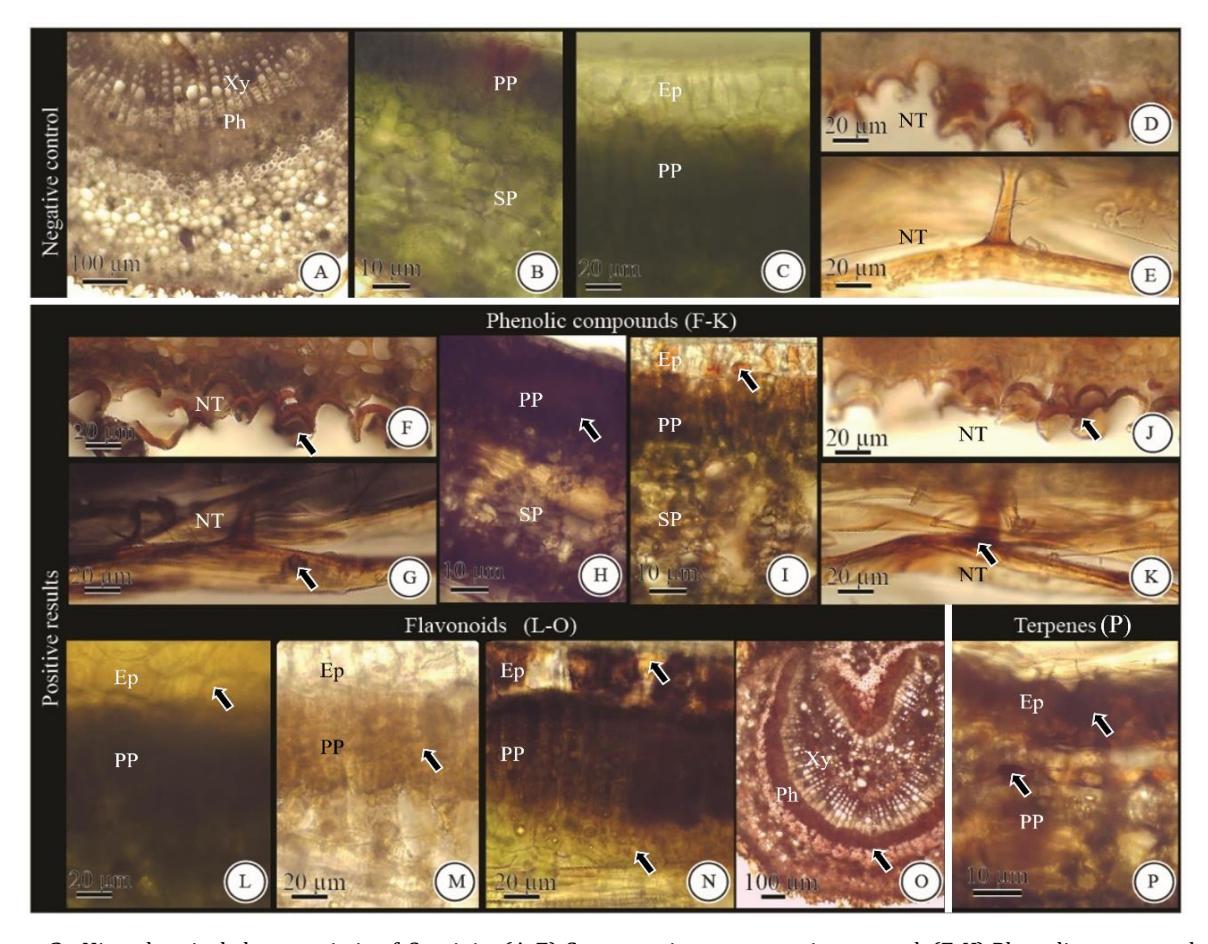
Figure 3. Histochemical characteristic of C. cainito (A-E) Cross-section as a negative control. (F-K) Phenolic compounds (F-H) dark brown with ferric chloride (I-K) brown with potassium dichromate. (L-O) Flavonoids stained (L-N) yellow with (L) aluminum chloride (M) citroboric acid (N) sodium hydroxide (O) pink with vanillin-HCl. (P) Terpenes stained brown with cupric acetate. Ep, epidermis; Ic, intercostal; Md, midrib; NT, non-glandular trichome; Ph, phloem; PP, palisade parenchyma; SP, spongy parenchyma; Xy, xylem. Arrows indicate idioblasts with chemical compounds.
In the transverse sections of M. zapota leaves (Figure 4A-D), without chemical treatment (negative control) was not possible to detect any chemical compounds. The histochemical tests showed that phenolic compounds are widely distributed in idioblast cells of intercostal (Figure 4E-F). The terpenes compounds were detected in the epidermal cells (Figure 4G) and the midrib (Fig. 4H). Flavonoids are located in idioblast cells of parenchyma, sponges in the midrib (Figure 4I), and intercostal area (Figure 4J-L). Islam et al. (2013) reported that flavonoids, tannins, saponins, and alkaloids were found in M. zapota leaves with phytochemical screening. The histochemical and phytochemical results of the alkaloids were different due to where the growing location of the plant. Previously, Moura et al. (2019) identified histochemical tests in the leaflets of M. zapota for phenolic compounds, and their results were similar to ours. These authors obtained positive tests with 10% potassium dichromate for phenolic compounds, vanillin hydrochloric acid for tannins, and antimony trichloride for triterpenes and steroids. Although this research used a different reagent for triterpenes and steroids, it showed positive results with similar localization. Phenolic compounds were identified using the same reagents with the same results. In this journal, there are different terms wherein vanillin is mentioned to detect tannins. According to Mace and Howell (1974), vanillin hydrochloric acid can identify tannin precursors, particularly
gallocatechin and catechin, as flavan-3-ol compounds. A study in the journal Guerin et al. (1971) also showed that vanillin hydrochloric acid could detect flavanol and flavane-diol compounds. Catechins were previously classified as tannins because they have similar ingredients (85% of them are the same). However, there are some differences, like the plants containing tannin can be used for tanning leather, but catechin does not have that function. Moreover, it is known that catechin and epicatechin polymerize tannins. (Rauf et al. 2019).
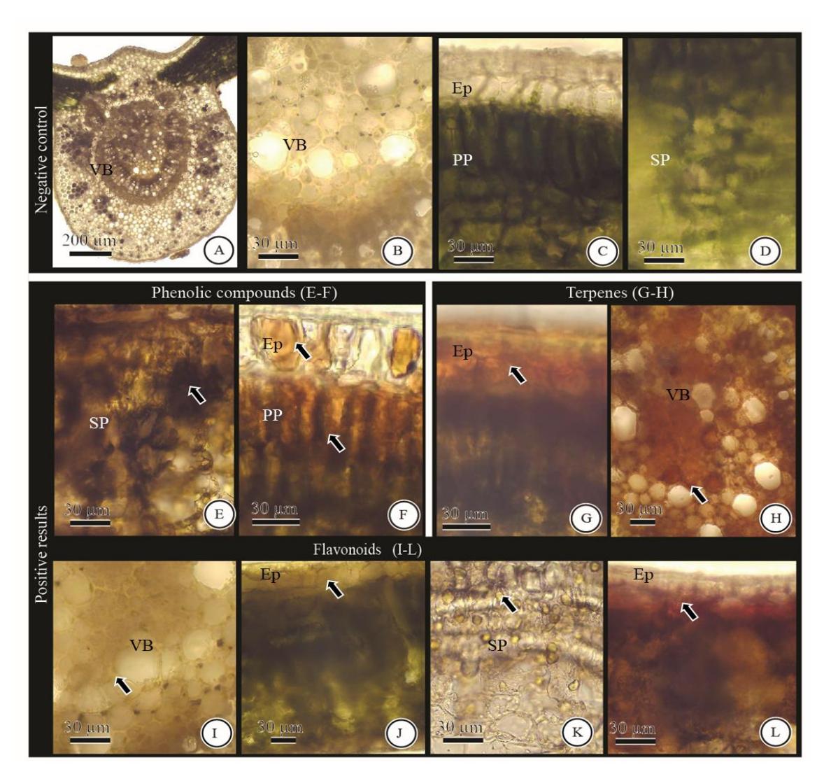
Figure 4. Histochemical characteristic of M. zapota (A-D) Cross-section as a negative control. (E-F) Phenolic compounds (E) dark brown with ferric chloride (F) brown with potassium dichromate. (G-H) Terpenes stained brown with cupric acetate. (I-L) Flavonoids stained (I-K) yellow with (I) aluminum chloride (J) citroboric acid (K) sodium hydroxide (L) pink with vanillin-HCl. Ep, epidermis; PP, palisade parenchyma; SP, spongy parenchyma; VB, vascular bundle. Arrows indicate idioblasts with chemical compounds.
The natural compounds, known as primary and secondary metabolites, are synthesized by secretory cells. The secretory cells occur in a single cell (idioblasts) or multicellular structures such as epidermis, hypodermis, parenchyma, trichomes, cavities, and ducts (Fahn 2000). Therefore, plants' secreting structure is found mainly in idioblasts (Mamoucha et al. 2016). The idioblasts can accumulate and secrete a compound such as oil, resin, mucilage, and tannin (Fahn 1979). During the evolution, secretory tissues seem to have developed from secretory idioblasts scattered among the normal tissues. Subsequently, ducts and cavities developed, and finally secretory trichomes (Fahn 1988). The internal secretory structures present a large distribution in four leaves, and the external secretory structures only present in D. kaki and C. cainito leaves. In V. varingiaefolium, the trichomes decrease and do not remain in some old stems, ripe fruit, and old leaves (Sholikhah et al. 2015).
The histochemical tests on four leaves of Ericales plants showed that phenolic compounds were presented in the idioblasts of the internal secretory structure. They can be detected using ferric chloride and potassium dichromate reagents, as demonstrated in other studies. Matias et al. (2016) performed histochemical tests on species of Solanum for phenolic compounds stained brown with ferric chloride and potassium dichromate reagents. Andrade et al. (2017), Betim et al. (2020) and Ribeiro et al. (2021) also showed that phenolic compounds reacted positively with ferric chloride (brown color). Besides the idioblasts, D. kaki, and C. cainito leaves also have trichomes that contained phenolic compounds. Phenolic compounds and glandular trichomes on D. kaki leaves also contain terpenes. The glandular trichomes often produce and accumulate terpenes and phenylpropanoid oils (Wagner et al. 2004). Another secondary metabolite accumulated in glandular trichomes was investigated histochemically as demonstrated by Liu and Liu (2012) on the leaves of Isodon rubescens (Lamiaceae) that contained terpenoids, flavonoids, carbohydrates, phenolics, and alkaloids. In the C. cainito leaves, the trichomes showed the location of phenolic compounds in nonglandular trichomes. The non-glandular trichomes, in this case, established the association with chemical reactions in the plants. Tozin et al. (2016)
also found that the non-glandular trichomes from Lamiaceae and Verbenaceae participate in the chemical interaction of the plants with the environment, supplementing the work of the typically glandular trichomes. It indicates that nonglandular trichomes functioned as physical and chemical protection in plants.
The flavonoid compounds were detected in the epidermis and parenchyma using aluminum chloride reagents, citroboric acid, and sodium hydroxide. Kuster and Vale (2016) studied four leaves of medicinal species from Cerrado and found the flavonoid location in parenchyma cells, especially in the palisade and spongy parenchyma, with histochemical tests. Different location of flavonoid in this study was shown with a vanillin-hydrochloric acid reagent which only showed positive reactions in idioblast cells of the midrib and intercostal area due to the limited detection capability of vanillinhydrochloric acid reagent to detect the presence of flavanol and flavandiol in plant tissues (Mace and Howell 1974, Guerin et al. 1971). In other cases, the aluminum chloride reagent detects flavonoids with free o-dihydroxy groups, while the citroboric acid and sodium hydroxide detect the presence of flavonoids in general (Pedro et al. 1990, Robinson 1983). According to Mulyani and Laksana (2011), the histochemical method can be used to analyze the presence of flavonoids in the test material, indicated by the yellow color with ammonia, NaOH, and AlCl3, citroboric acid reagents. Mamoucha et al.(2016) used vanillin-HCl to observe flavonoid compounds that showed pink color in the leaf of Ficus carica. These two studies have the same occurred in our study.
CONCLUSION
The histochemical test is a useful technique for detecting bioactive substances in secretory medicinal plants. In contrast to the conventional phytochemical screening procedure, only a crosssection sample and a few drops of the reagent are required. The four leaves contained phenolic compounds, flavonoids, and terpenes, while quinones were only detected in cantigi leaves. Internal secretory structures were observed in the midrib (leaf bone) and intercostal (part of the leaf bone and leaf edges), especially as idioblast cells. The idioblast cells can be seen on the epidermis, parenchyma, and vascular tissues. Meanwhile, the external secretory structures in trichomes containing phenolic compounds were only found in D. kaki, and C. cainito leaves. Besides phenolic compounds, the glandular trichomes in D. kaki leaves also had terpenes.
ACKNOWLEDGMENTS
None. No funding to declare
REFERENCES
Andradea EA, Folquitto DG, Luzc LEC, Paludo KS, Farago PV, Budel JM, 2017, Anatomy and histochemistry of leaves and stems of Sapium glandulosum, Rev bras farmacogn 27: 282-289.
APG IV, 2016, An update of the angiosperm phylogeny group classification for the orders and families of flowering plants: APG IV. Bot J Linn Soc. 181:1Ȃ20.
Barbosa SM, Abreu N do C, Oliveira MS de, Cruz JN, Andrade EH de A, Neto MAM, Gurgel ESC, 2021, Effects of light intensity on the anatomical structure, secretory structures, histochemistry and essential oil composition of Aeollanthus suaveolens Mart. ex Spreng. (Lamiaceae), Biochem. Syst. Ecol. 95: 1-11.
Betim FCM, Barbosa V, Oliveira CF de, Miguel OG, Raman V, Budel JM, Miguel MD, Dias J de FG, 2020, Microscopy and histochemistry of Ocotea nutans (Nees) Mez (Lauraceae), Flora 273: 1-6.
Coelho VPM, Leite JPV, Nunes LG, Ventrella MC, 2012, Anatomy, histochemistry and phytochemical profile of leaf and stem bark of Bathysa cuspidata (Rubiaceae), Aust J Bot 60: 49-60.
Coleman R, 2000, The impact of histochemistry - a historical perspective, Acta histochem. 102: 5-14.
Dewick PM, 2009, Medicinal Natural Products: A Biosynthetic Approach, 3rd edn, John Wiley & Sons, United Kingdom.
Dickison WC, 2000, Integrative plant anatomy, Academic Press, New York.
Fahn A, 1979, Secretory tissues in plants. London: Academic Press.
Fahn A, 1988, Secretory tissues in vascular plants, New Phytol 108: 229-257.
Fahn A, 2000, Structure and function of secretory cells. In: Hallahan DL, Gray JS (eds) Plant trichomes. Advances in botanical research, Vol. 31, Academic Press, London.
Farnsworth NR, 1966, Biological and Phytochemical Screening of Plants, J Pharm Sci 55(3): 243-268.
Furr M, Mahlberg PG, 1981, Histochemical analyses of laticifers and glandular trichomes in Cannabis sativa, J Nat Prod 44: 153-159.
Gabe M, 1968, Techniques histologiques, Masson, Paris.
Ganguly A, Rahman SMA, 2013, Pharmacological Studies of Leaves of Manilkara Zapota (Sapotaceae), Extensive Invivo and In-vitro studies. Lambert Academic Publishing, Germany.
Gomez AA, Mercado MI, Belizán MME, Ponessa G, Vattuone MA, Sampietro DA, 2019, In situ histochemical localization of alkaloids in leaves and pods of Prosopis ruscifolia, Flora 256: 1-6.
Guerin HP, Delaveau PG, Paris RR, 1971, Localisations histochimiques.: II: Procédés simples de localisation de pigments flavoniques. Application à quelques Phanérogames. Bulletin de la Société Botanique de France 118: 29-36.
Islam MR, Parvin MS, Banu MR, Jahan N, Das N, Islam ME, 2013, Antibacterial and phytochemical screening of ethanol extracts of Manilkara zapota leaves and bark, Int J Pharma Sci 3(6): 39-397.
Johansen DA, 1940, Plant Microtechnique, McGraw-Hill, New York.
Kuster VC, Vale FHA, 2016, Leaf Histochemistry Analysis of Four Medicinal Species from Cerrado, Rev Bras Farmacogn 26: 673-678.
Lavis LD, 2011, Histochemistry: live and in color. J Histochem Cytochem 59(2):139-145.
Liu M, Liu J, 2012, Structure and histochemistry of the glandular trichomes on the leaves of Isodon rubescens (Lamiaceae), Afr. J. Biotechnol 11(17): 4069-4078.
Mace ME, Howell CR, 1974, Histochemistry and identification of condensed tannin precursor in roots of cotton seedlings, Can J Bot 52: 2423-2426.
Mamouchaa S, Fokialakisb N, Christodoulakisa NS, 2016, Leaf structure and histochemistry of Ficus carica (Moraceae), the fig tree, Flora 218: 1-11.
Matias MJ, Mercadante-Simoes MO, Royo VA, Ribeiro LM, Santos AC, Fonseca JMS, 2016, Structure and histochemistry of medicinal species of Solanum, Rev bras farmacogn 26: 147-160.
Mercadante-Simoes MO, Mazzottini-Dos-Santos HC, Nery LA, Ferreira PRB, Ribeiro LM, Royo VA, Oliveira DA de, 2014, Structure, histochemistry and phytochemical profile of the bark of the sobol and aerial stem of Tontelea micrantha (Celastraceae - Hippocrateoideae). An Acad Bras Cienc 86 (3): 1167- 1179.
Moura BI de V, Araújo BPL de, Sá RD, Randau KP, 2019, Pharmacobotanical study of Manilkara zapota (L.) P.Royen (Sapotaceae). Braz J Pharm Sci 55, e17227.
Mulyani S, Laksana T, 2011, Analisis Flavonoid Dan Tannin dengan Metoda Mikroskopi mikrokimiawi. Majalah Obat Tradisional 16(3): 109-114.
Pedro L, Campos P, Pais M, 1990, Morphology, ontogeny and histochemistry of secretory trichomes of Geranium robertianum (Geraniaceae). Nordic Journal of Botany 10(5): 501-509.
Ribeiro OD, Nascimento WMO do, Cruz FJR, Gurgel ESC, 2021, Seed anatomy and histochemistry of Myrciaria dubia (Kunth) McVaugh, an Amazonian Myrtaceace. Flora 280: 1-7.
Robinson T, 1983, The organic Constituents of Higher Plants Their Chemistry and Interrelationships, 5th edn, Cordus Press, North Amherst.
Rocha M, Figueiredo MR, Kaplan MAC, Durst T, 2015, Chemotaxonomy of the Ericales. Biochem Syst Ecol 61: 441-449.
Royo V de A, Mercadante-Simões MO, Ribeiro LM, Oliveira DA de, Aguiar MMR, Ellenhise RC, Ferreira PRB, 2015, Anatomy, Histochemistry, and Antifungal Activity of Anacardium humile (Anacardiaceae) Leaf. Microsc. Microanal: 1-13.
Rupa D, 2015, Identification of Secretory Structure, Histochemical and Phytochemical Analysis of Anti-
Infection Medicinal Plants in the Bukit Duabelas National Park of Jambi. Tesis. Institut Pertanian Bogor, Bogor.
Rauf A, Imran M, Abu-Izneid T, Iahtisham Ul H, Patel S, Pan X, et al., 2019, Proanthocyanidins: a comprehensive review Biomed. Pharmacother, 116, p. 108999
Shailajan S, Gurjar D, 2014, Pharmacognostic and Phytochemical Evaluation of Chrysophyllum cainito Linn. Leaves, Int J Pharm Sci Rev Res 26(1): 106-111.
Sholikhah A, Dian FA, Listyorini D, 2015, Anatomy and Morphological Study of Mentigi Gunung (Vaccinium varingiaefolium (Blume) Miq.) in Area of Mount Batok-Indonesia, Proceeding of international conference on biological science, KnE Life Sciences, pages 36Ȃ45.
Tozin LRS, Silva SCM, Rodrigues TM, 2016, Nonglandular trichomes in Lamiaceae and Verbenaceae species: morphological and histochemical features indicate more than physical protection, N Z J Bot, 54(4): 446-457.
Xie C, Xie Z, Xu X, Yang D, 2015, Persimmon (Diospyros kaki L.) leaves: A review on traditional uses, phytochemistry and pharmacological properties, J Ethnopharmacol163: 229-240.
Wagner GJ, Wang E, Shepherd RW, 2004, New approaches for studying and exploting an old protuberence, the plant trichome, Annals of Botany 93: 3-11.
Yang L, Wen KS, Ruan X, Zhao YX, Wei F, Wang Q, 2018, Response of plant secondary metabolites to environmental factors, Molecules 23(4): 1Ȃ26.
Yulyana A, Winarno H, Kosasih, 2016, Karakterisasi Ekstrak Daun Cantigi (Vaccinium varingiaefolium Miq.), J Sains Kes 1(5).
Zuhro F, Puspitasari E, Muslichah S, Hidayat MA, ʹͲͳ, Aktivitas Inhibitor Ƚ-Glukosidase Ekstrak Etanol Daun Kenitu (Chrysophyllum cainito L.), Pustaka Kesehatan 4(1): 1-7.
Zreen Z, Kiran S, Hameed A, Gulzar T, Farooq T, 2019, Phytochemical and comparative biological studies of Diospyros kaki extracts, Int. J. Biosci. 14(1): 207-223.