

INTRODUCTION
Single crystals are very important in modern metallurgical research. These crystals are used as specimens for X-ray study and as specimens for research in understanding metal behaviour during deformation. The use of single crystals in the study of deformation behaviour simplifies the calculation, since in this case the effect of grain boundaries and grain orientation differences are eliminated.
Before this work, only one method of growing \(Ag_3Sn\) single crystals was known. This method was developed by Fairhurst and Cohen (1) and produced \(Ag_3Sn\) single crystals in granular form. These crystals have proven their superiority for analising their crystal structure but fail for studying their deformation behaviour. Wirjosumarto (2) in his study on the deformation mechanism of \(Ag_3Sn\), has shown that platelet single crystals were more suitable than the granular ones.
In this paper, the method of growing and the method of orientation determination for platelet Ag<sub>3</sub>Sn single crystals are explained and discussed.
MASERIAL AND METHOD
Material
The chemical composition of Ag<sub>3</sub>Sn is 73.15 wt % of silver and 26.85 wt % of tin. To produce platelet single crystals of this compound, however, ingots of 40 wt % silver and 60 wt % tin were used. These ingots were prepared from silver and tin of 99.99% purity.
2. Experimental Method
a. Method for Preparing the Ingot
The ingot was prepared by melting a mixture of 40-60 wt % of silver and tin in a Vycor reaction glass tube with inner diameter of 5 mm. The ingot was then homogenized at 400°C for 48 hours. During the homogenization process the ingot was sealed in an evacuated glass tube to minimize oxydation.
b. Method for Growing the Crystal
The method used in this work was a temperature gradient method. It is a modification of Bridgman's (3) technique. This method consists of remelting the homogenized ingot in a Vycor reaction glass tube and cooling it to solidify; then followed by passing the reaction glass with its content through a 500°C furnace at a speed of 1.5 mm per hour. The gradient solidified ingot was then immersed in concentrated hydrochloric acid for at least 3 days in order to dissolve the eutectic constituent.
The microstructure of the ingot, before it was immersed in concentrated hydrochloric acid, was photographed. The preparation of the surface for the metallographic study was done according to the standard method and followed by etching it in a modified Crowell's etchant*).
c. Method for Determining Crystal Orientation
An X-ray diffraction pattern of the surface of a platelet single crystal was taken with a General Electric X-ray diffraction unit using Cu-K\(\alpha\) radiation. This pattern was then compared to that of polycrystalline Ag<sub>3</sub>Sn powder in order to prove that it is a single crystal.
In order to determine precisely the crystallographic plane of the single crystal surface, a more accurate X - ray diffraction method was employed. This was done by using step scanning method on the diffraction peak of the single crystal surface and on the corresponding diffraction peak of the polycrystalline powder sample. The step scanning was done on Picker X-ray diffraction unit with intervals of 1/60 degree and irradiation of one minute for each step with Cu-K\(\beta\) radiation.
Full orientation of the crystal was determined by a scanning search made at each Bragg angle. This was done on a Norelco X-ray diffraction unit by employing pole figure device.
RESULTS AND DISCUSSIONS
a. Microstructure of the Gradient Cooled Ingot
The microstructure of the ingot after the gradient cooling is shown in Figure 1. The micrograph shows that the ingot consists of a layer structure. This structure was expected, since the composition of the ingot was 40 wt % silver and 60 wt % tin. This composition at room temperature consists of Ag_Sn or \(\gamma\) phase and the eutectic composition of Ag_Sn alloy. A composition of 40-60 wt % Ag_Sn was chosen in order to ensure that during the solidification process, only solid \(\gamma\) and solid eutectic are produced. These are explained by the binary phase diagram of silver and tin which is shown in Figure 2.
*) K<sub>2</sub>Cr<sub>2</sub>O<sub>7</sub> 9 grams. H<sub>2</sub>SO<sub>4</sub> 36 cc. NaF (sat. solution) 18 cc. H<sub>2</sub>O 450 cc.
light = \(Ag_3Sn\) or \(\gamma\); dark = eutectic
Figure 1. Microstructure of a gradient cooled 40-60 wt % Ag-Sn ingot, etched with modified Crowell's etchant
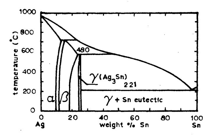
Figure 2. Phase diagram of Ag-Sn system
I a e TI au pu pe c1
de:
th
By fast cooling the \(\gamma\) phase forms granular structure. By gradient cooling however, layer structure of \(\gamma\) was formed. This process is explained in the following.
The solidification temperature of \(\gamma\) (480°C) is higher than that of the Ag-Sn eutectic (221°C). So during the gradient cooling, \(\gamma\) or \(\text{Ag}_3\text{Sn}\) solidifies first and grows into the molten metal by absorbing Ag from the liquid to form \(\text{Ag}_3\text{Sn}\). In this way the silver concentration of the molten metal becomes lower and lower until eutectic composition is reached. When this composition has been reached and when the solidus line of 221°C is passed, then the molten eutectic metal which fills the space between the solid platelet of \(\text{Ag}_3\text{Sn}\), solidifies simultaniously. This solidification process produced the mentioned layer structure and after dissolving the tin eutectic in concentrated hydrochloric acid, platelets of \(\text{Ag}_3\text{Sn}\) with thickness of about 200 \(\mu\text{m}\) and about 5 mm in other dimensions were obtained.
b. Proof and Orientation of the Single Crystals
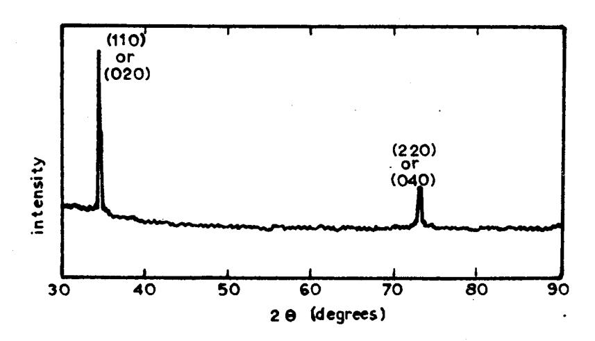
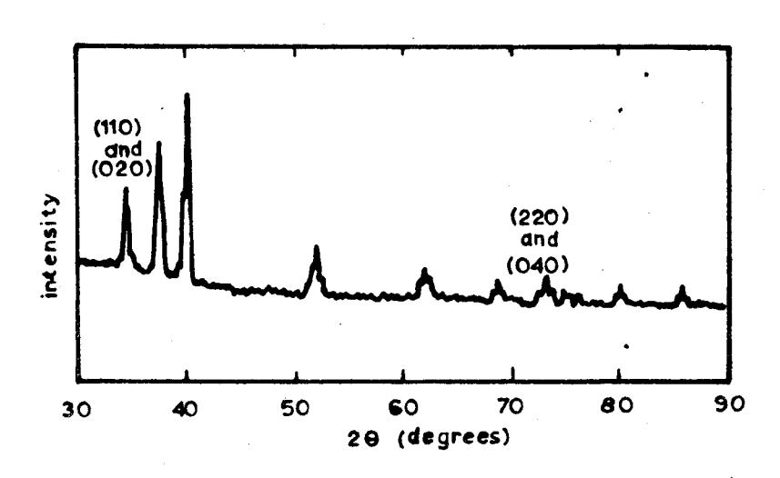
The result of the X-ray diffraction of the platelet \({\rm Ag}_3{\rm Sn}\) is shown in Figure 3a and that of the polycrystalline powder in Figure 3b. By comparing these two patterns, it can be deduced that the surface of the platelet \(\gamma\) is either (110) plane or (020) or both (110) and (020) planes. Based on this there exists 4 possibilities, they are:
- 1. The platelets are single crystals with (110) face,
- 2. They are single crystals with (020) face,
- 3. Polycrystalline with two orientations, and
- 4. They are bicrystals.
In order to be able to evaluate correctly, the results of the step scanning X-ray diffraction of both platelet \({\rm Ag}_3{\rm Sn}\) and polycrystalline powder of the same X-ray peak, which is shown in Figure 4, were analyzed. This X-ray method is able to separate several closely spaced peaks and the use of Kß radiation eliminates the complication caused by \({\rm K}\alpha_1\) and \({\rm K}\alpha_2\) doublet. These patterns show the peakprofile of both samples at Bragg angle corresponding to the (110) and (020) peaks. From these profiles it can be seen clearly that the platelet has only one peak and it corresponds to the (020) peak. So it can be concluded that the platelets are single crystals of \({\rm Ag}_3{\rm Sn}\) and their flat surfaces are (020) plane.
Complete orientation determination of the single crystals were done with X-ray diffraction method employing pole figure device. The result is shown in Figure 5. The complete orien-
a. Platelet of Ag<sub>3</sub>Sn or γ
b. Polycrystalline powder
Figure 3. X-ray diffraction patterns of platelet Ag<sub>3</sub>Sn (a) and polycrystalline powder (b) with Cu-Ka radiation
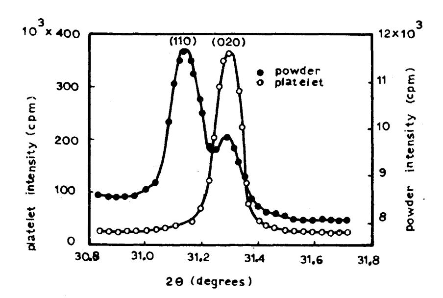
Figure 4. X-ray peak profiles of platelet and powder samples with Cu-K\(\beta\) radiation
Figure 5. Surface micrograph and principle axis directions of \({\rm Ag_3Sn}\) platelet single crystal (unetched)
tation of the crystal is then, the \(\bar{b}\) axis or the <010> direction is the normal of the flat surface and the <100> direction or \(\bar{a}\) axis and <001> or \(\bar{c}\) axis are as shown in the figure.
CONCLUSIONS
- Platelet single crystals of Ag<sub>3</sub>Sn can be produced from an ingot of 40 wt % Ag and 60 wt % Sn using modified Bridgman's technique.
- 2. The surfaces of the platelet single crystals produced in this way have the same crystallographic plane, they are (020) plane.