

Sari
Pengaruh suhu substrat pada film tipis 'disperse red 1' yang dideposisi pada gelas ITO dalam vakum
Dalam eksperimen ini telah berhasil diperoleh melalui proses deposisi vakum sejumlah film tipis bahan fotorefraktif dari molekul "Disperse Red 1" (DR1) dengan kristalinitas tinggi di atas substrat gelas ITO ("indium tin oxide") pada berbagai suhu. Selain efek orientasi dan organisasi molekul yang ditunjukkan oleh karakteristik spektra XRD dan FTIR bersangkutan serta peningkatan efek tersebut dengan suhu substrat, analisis lebih lanjut dari data FTIR di sekitar pita absorpsi nitrobenzena telah mengungkapkan tanda pembentukan ikatan hidrogen yang kuat yang menghasilkan susunan "head-tail" yang bertumpuk. Film yang terdeposisi juga memperlihatkan pengurangan absorpsi cahaya secara sistematik di daerah tampak, seiring dengan peningkatan suhu substrat. Perubahan sifat optik ini jelas bermanfaat bagi aplikasi film DR1 untuk devais fotonik dalam daerah gelombang panjang.
Kata kunci: deposisi vakum; "disperse red 1"; film tipis; sifat optik suhu substrat.
1 Introduction
Optical nonliniear (ONL) organic molecules consisiting of conjugated chain and electron donor as well as electron acceptor groups such as DANS, MONS, DO3 and DR1, have been studied for their photorefractive and second order optical nonlinear properties which are useful for a variety of applications such as electrooptic modulation, optical switching, frequency doubling, and optical data storage [1-7]. For those photonic device applications, the ONL organic materials are generally required to be prepared in the form of good quality thin film, characterized by its good optical transparancy, thickness homogeneity and surface smoothness in addition to its appropriate nonlinear optical properties.
The physical vacuum deposition (PVD) method has been demonstrated recently as a viable alternative for organic thin film fabrication, specifically for low molecular weight chromophores molecules such as p-NA [8], DMANS [9] and Disperse Red 1 [10, 11]. In addition to producing film with high purity and homogeneity, this method has also proved to be effective in promoting molecular orientation in the film [8-11]. This effect is expected to induce changes in the film's optical property, and with further arrangement of the oriented molecules, will most likely lead to considerable enhancement of its photorefractive and nonlinear optical properties.
In our previous report on the result of physical vacuum deposition of DR1 film on slide glasses [10, 11], the molecules in the film were shown to be organized and oriented perpendicular to the film surfaces. In this experiment, the PVD method was employed for the preparation of Disperse Red 1 (DR1) thin film on ITO glass substrate at substrate temperatures in the same range of variation. The different substrate temperatures will be shown to induce changes in the optical absorption characteristic of the resulted films. This result will be compared with our previous result on DR1 thin film vacuum deposited on slide glass substrate [11].
2 Experimental
2.1 Material
Powder of Disperse Red 1 (4-{N-ethyl-N-(2-hydroxyethyl)}amino-4'-nitroazobenzene ) was obtained commercially from Sigma. This molecule has a formula weight of 314.3, and the measured values of melting point and the onset temperature of thermal decomposition are 153°C and 219°C, respectively [11]. The structure of the molecule is shown in Figure (1). This powder was used for vacuum deposition without further treatment. As seen from the figure, this molecule consists of {N-ethyl-N-2-ethylhydroxy} amino as the electron donor group, and NO<sub>2</sub> as the electron acceptor groups, with the conjugated-chain azobenzene serving as the \(\pi\)-electron bridge between the two groups.
Figure 1 Molecular structure of DR1 [12]
2.2 Sample preparation
Disperse Red 1 thin film was prepared in VFC-410 Vacuum Evaporator from Ulvac Sinku Kiko, which was operated at about 3 x 10<sup>-5</sup> Torr. The Disperse Red 1 (DR1) thin films were deposited on clean microscope slides glass, and ITO glass. For the deposition processes these substrates were placed at about 10 cm above the crucible. Following our previous experimental result on the optimal evaporation temperature for the deposition process [11], the crucible temperature was set at 173°C in this experiment, while the setting of substrate temperature was more or less equally spaced at 26°C, 55°C and 75°C. Above 75°C, the deposition rate dropped drastically due to re-evaporation effect. No further annealing treatment was conducted on the as-grown films.
2.3 Characterization
The crystallinity and crystal structure were examined on the basis of XRD patterns obtained by Philips Diffractometer which was operated with the CuK\(\alpha\) (\(\lambda\) = 1.5406 Angstrom) X-Ray source. The optical measurement of the pristine polycrystalline powder and vacuum deposited DR1 thin film were characterized by a Beckman DU-7000 Single Beam Spectrophotometer. The thickness of the film was determined by Dektak IIA depth profiler.
3 Results and discussions
The measured thickness of the evaporated film varies from 0.3 to 0.7 µm for different films. The deposited
films generally exhibit smooth surface and homogeneous thickness.
The result of FTIR measurement presented in Fig. (2) consists of spectrum of the (a) pristine DR1 powder, (b) DR1 powder obtained by pulverizing the film deposited at substrate temperature of 26°C, and that of (c) the deposited film. It is observed that there is no meaningful difference between spectrum (a) and (b), implying no significant structural change due to the deposition process. Meanwhile, the presence of molecular orientation effect is clearly indicated by comparison of (a) with (c) as discussed in detail in our earlier work [11].
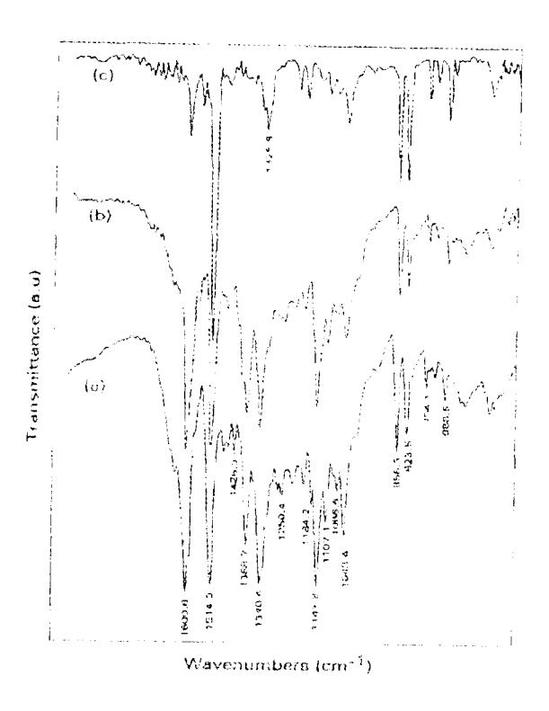
Figure 2 FTIR spectra for samples of (a) pristine powder, (b) powder pulverized from deposited film, and (c) film as deposited on Si substrate.
Figure (3) shows the results of XRD measurement for thin film as deposited on ITO glass at substrate temperatures of 26°C (a), 55°C (b), and 75°C (c), respectively. This figure was normalized to the film's thickness. The XRD pattern of ITO glass (without DR1 film) is also presented along in this figure for comparison and proper interpretation of the spectra. The diffraction peak at \(2\theta = 9^{\circ}\) corresponds to interlayer distance (d) of 9.8 Å which is not far from the length of azobenzene in the trans-isomer of DR1 (~ 9 Å) [13] suggesting that DR1 molecules are oriented perpendicular to the substrate's surface in a head-tail configuration as suggested in our previous work [11]. The peak at \(2\theta =\)30° originated from the ITO glass remains more or less constant with respect to the glass temperature. While the peak intensity associated with DR1 thin film does not
seem to undergo any visible change, but the spectral width does display visible reduction with increasing substrate temperature as observed in Fig. 3(b), (c) and (d). This systematic change of the XRD spectrum indicates an improved crystallinity (crystalline molecular organization) in the film with increasing substrate temperature.
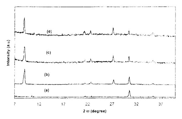
Figure 3 XRD patterns of (a). ITO glass without DR1 film, (b). Disperse Red 1 thin film deposited on ITO glass at 26°C, (c) 55°C, (d). 75°C.
In connection with the suggested head-tail stacking of those molecules it is useful to take a closer look at Fig. (2), specifically around the nitrobenzene band at 1340 cm<sup>-1</sup>, which has been scaled up in Fig. (4) for a close-up view. Even a cursory view on those spectral bands will lead to a confirmation of the formation of the head-tail configuration via the strong hydrogen bonding of the type O-H "O between the nitro group of the acceptor and the hydroxyl group in the donor as depicted in Fig. (5) and explained in the following. We note first of all a visible reduction of the 1340 cm<sup>-1</sup> absorption band going from (a) to (b), with the appearance of a small kink to its right (located approximately at 1330 cm<sup>-1</sup>). It is to be expected on general ground that the formation of hydrogen bonding will suppress the above mentioned absorption band. However, the suppression is less than complete since a good part of these bonds were already disrupted by the process of pulverization. The presence of the remaining bonds responsible for the reduction of the 1340 cm<sup>-1</sup> band is also confirmed by the appearance of the small kink to its right as alluded above. This interpretation is further evidenced by the near disappearence of the 1340 cm<sup>-1</sup> band along with the strong enhancement of the 1329 cm<sup>-1</sup> band shown in Fig.4 (c) which is associated with the effect of hydrogen bonding induced by the deposition process. It is well known that the formation of hydrogen bonding [14] will generally lead to a down shift of the absorption band along with its broadening, in conformity with the observation described above. It is further noted that the enhanced intensity of the 1329 cm<sup>-1</sup> band is still short of that of the original 1340 cm<sup>-1</sup> band since the aligned molecules are expected to suppress the contribution coming from the vibration mode along the long molecular axis.
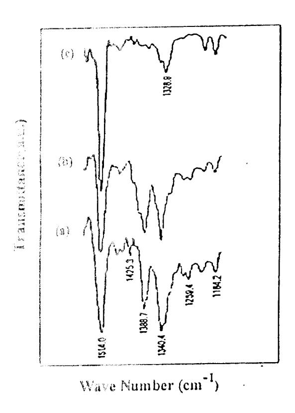
Figure 4. A close-up view of FTIR spectra around the nitro pennent of absorption band for samples of (a) pristine powder, (b) powder politerized from deposited film, and (c) film as deposited on Figure train.
Figure 7. The form of hydrogen bonding between the nitro group and that it arrange group of two DR1 molecules as suggested by Eq. (4).
PROC. ITB. VOL. 33, NO. 1, 2001
Figure (6) shows the results of the UV-VIS measurements on the solution of pristine Disperse Red 1 powder (A) and solution of Disperse Red 1 powder pulverized from film deposited at 26°C (B), both dissolved in NMP solvent. This figure shows a broad and practically identical absorption band in the visible region with a wavelength of maximum absorption located at 508 nm (\(\lambda_{\text{max}}\)=508 nm). This broad absorption band in VIS-region (508 nm) is commonly attributed to intramolecular charge transfer (CT) transition of \(\pi\)electron from donor (N-ethyl-2N-ethylhydroxyl) to acceptor group (NO<sub>2</sub>) via the conjugated-chain azobenzene [15]. The identical appearance of the two absorption bands in this region implies that thermal deposition process did not cause any change in the conjugated structure of DR1 molecule. The slight shift of the peak together with the considerable suppression of the UV band of the pulverized film may also be related to the presence of the remaining head-tail hydrogen bonds in the sample. However, the detailed explanation of the changes requires further experimental study.
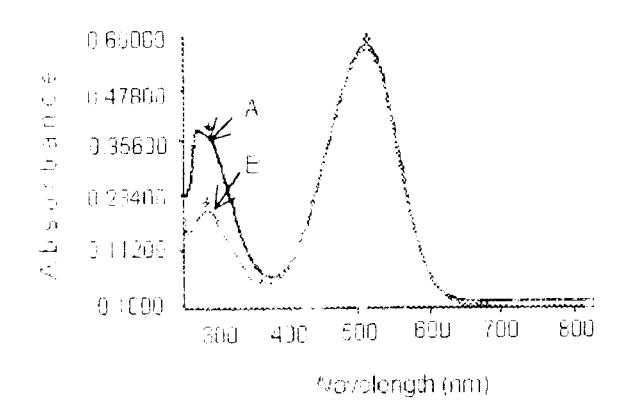
Figure 6 UV-VIS spectra for (A) solution of Disperse Red 1 pristine powder in NMP (B) solution of Disperse Red 1 powder pulverized from film deposited at 26°C.
The effect of substrate temperature on the UV-VIS absorption spectra of thin films deposited on ITO glass at 26°C, 55°C and 75°C is described in Fig.7. It is clear that new features appear in these spectra. But a most remarkable effect in this spectra is the pronounced reduction of the long-wavelength (visible) absorption band. This effect is considerably larger than that observed in the film vacuum deposited on slide glass [11]. It is certainly a welcome change favorable for the development of long wavelength applications of photonic device. Another interesting change induced by the deposition process at higher substrate temperature is the appearance and enhancement of a new band at the local n-π* transition of trans azobenzene [16], a molecular configuration favored at higher temperature. Fig.7 also displays an intriguing effect manifested in the enhancement of the UV absorption band, its split structure and a relative intensity shift in favor of its lower wavelength component. We note that qualitatively
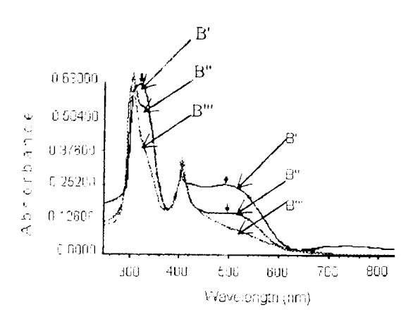
Figure 7 UV-VIS spectra of Disperse Red 1 thin films as deposited on ITO glass substrate at (B') 26°C (B") 55°C (B"') 75°C, indicating modification of optical properties induced by elevated substrate temperature.
similar characteristic changes due to higher substrate temperature were also observed in film deposited on slide glass as shown in Fig.8 [11] for comparison. It must be stressed however, that the changes observed in the two cases differ in detail as well as quantitatively. A consistent explanation for the two cases is unfortunately not available from this experimental result.
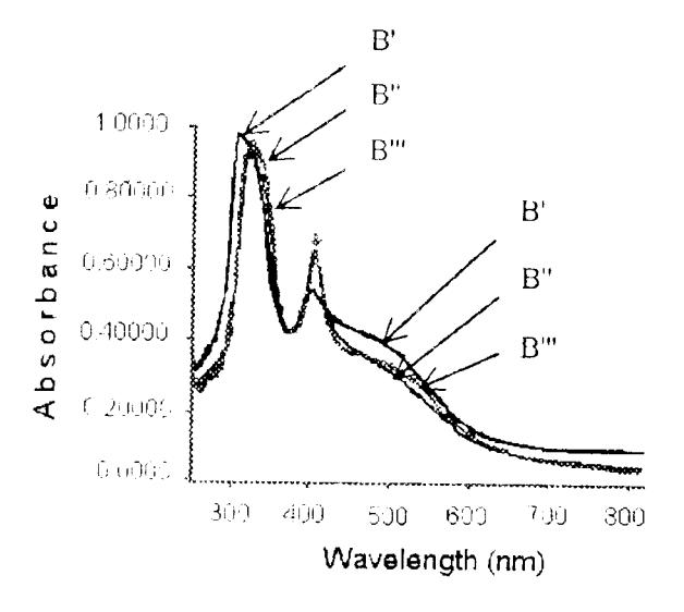
Figure 8 UV-VIS spectra of Disperse Red 1 thin film as deposited on glass substrate at (B') 26°C (B") 55°C (B"') 75°C
4 Conclusions
It has been demonstrated in this experiment that homogeneous thin film of DR1 could be vacuum deposited on ITO substrate. It was shown that the film exhibits similar spectroscopic features indicating molecular orientation and organization effects as shown by the films deposited previously on a slide glass substrate. We further substantiated the proposed head-tail molecular stacking organization by indicating the role of hydrogen bonding from the FTIR data. Among the most remarkable effect observed in this experiment in the long wavelength absorption reduction at elevated susbtrate temperature, which promised further development of DR1 film for photonic device applications.
5 Acknowledgments
We thank Mr. Aminudin Sulaeman for help with FTIR and UV-VIS measurements.
6 References
- 1. Prasad, PN, Orczyk, ME, & Zieba, J, Polymeric Composite Materials for Nonlinear Optics and Photonic, Nonlinear Optics, 14, pp 127-134, 1995.
- 2. Kaino, T, Yakoo, A, Asabe, M, Tomaru, S, & Kurihara, T, Organic Nonlinear Optical Materials for Device Applications, Nonlinear Optics, 14, pp. 135-150, 1995.
- 3. Mohlmann, GR, Development of Optically Nonlinear Polymers and Devices, Synthetic Metals, 37, 207-221, 1990.
- 4. Hu, S, Carlisle, GO, & Martinez, DR, Absorption Studies of polymer dye film oriented by corona poling, Journal of Materials Science Letter, 11, 994-796, 1992.
- Sotyama, W. Tatsuura, S. Motoyoshi, K. & Yoshimura, T. Directional-Coupled Optical Switch Between Stacked Waveguide Layers Using Electro-Optic Polymer, Jpn. J. Appl. Phys., 31, 1180-1181, 1992.
- Shishido, A, Tsutsumi, O, Kanazawa, A, Shiono, T, Ikeda, T, & Tamai, N, Rapid Optical Switching by Means of Photoinduced Change in Refractive Index of Azobenzene Liquid Crystals Detected by Reflection-Mode Analysis, J. Am. Chem. Soc., 119, 7791-7796, 1997.
- 7. Meng, X, Natanshon, A, Barrett, C, & Rochon, P, Azo Polymers for Reversible Optical Storage. 10.
- Cooperative Motion of Polar Side Groups in Amorphous Polymers, Macromolecules., 29, 946-952, 1996.
- 8. Okamoto, N, Unoh, H, Sugihara, O, & Matsushima, R, Large Second-Harmonic Generation in Composite Film of p-Nitroanilin and Its Derivatives, Nonlinear Optics., 14, 245-250, 1995.
- 9. Ehara, T, Hirose, H, Kobayashi, H, Kotani, M, Molecular alignment in organic thin films, Synthetic Metals, 109, 43-46, 2000.
- Taunaumang, H, Herman, Tjia, MO, Preparation of Disperse Red 1 Thin Film by Vacuum Evaporation Method, Prosiding Simposium Fisika Nasional XVIII, Serpong, 465-472, 2000.
- 11. Taunaumang, H, Herman, Tjia, MO, Molecular Orientation in Disperse Red 1 Thin Film Produced by PVD Method, subsmitted to Optical Material.
- 12. Singer, KD, Sohn, JE, & Lalama, SJ. Second harmonic generation in poled polymer films, Appl. Phys. Lett., 49, (5), 1986.
- 13. Nuyken, O, Scherer, C, Baindl, A, Brenner, AR, Dahn, U, Gartner, R, Kaiser, S, Matusche, P, & Voit, B, Azo-Group-Containing Polymers for Use in Communication Technologies, Prog. Polym. Sci., 22, 93-183, 1997.
- 14. Colthup, NB, Daly, LH, Wiberly, SE, Introduction to Infrared and Raman Spectroscopy, Academic Press, London, 1975.
- 15. Sotoyama, W, Tatsuura, S, & Yoshira, T, Electrooptic Side-Chain Polyimide System with Large Optical Nonlinearity and High Thermal Stability, Appl. Phy. Lett., 64, (17), 1994.
- Åstrand, P-O, Ramanujam, PS, Hvilsted, S, Bak, KL, & Sauer, SPA, Ab Initio Calculation of the Electronic Spectrum of Azobenzene Dyes and Its Impact on the Design of Optical Data Storage Materials, J. Am. Chem. Soc., 122, 3482-3487, 2000.