
1 Introduction
Zinc oxide (ZnO) has a direct energy band gap of 3.4 eV, which is useful for multiple applications, such as transparent conductive oxide coatings, sensors for various gases, biosensors and display panel technology. For the growth of various nanoarchitecture morphologies of ZnO nanomaterial, it is necessary to set the preparation parameters that affect the physical properties of the different morphologies. The ZnO nanostructure can be synthesized by various methods, such as thermal evaporation [1], sputtering [2], and chemical vapor deposition [3]. In view of the complications of methods with a high growth temperature, high preparation costs and the need of high vacuum conditions, chemical methods, such as hydrothermal synthesis [4] and aqueous solution [5], are preferrable for the fabrication of ZnO nanorods. In 2011, Amin, et al. [6] investigated the effect of pH value, precursor concentration, growth time and temperature on the morphology of ZnO nanostructures. They found a linear relationship between the diameter and the length of the ZnO nanorods with time up to 10 hours. Above this range no further increase was found. Meanwhile, Ghayour, et al. [7] showed the effect of a sputtered seed layer on the crystal structure, grain size and surface roughness of ZnO nanorods. They found that the crystallinity, grain size, diameter and alignment of the nanorods improved as the thickness of the seed layer was increased, while the roughness decreased. In 2013, Xian, et al. [8] showed that the diameter size of nanorods from 30 to 200 nm can be controlled via seed layer annealing and that the ZnO nanorods change from a needle- to a hexagonal-like structure. In 2012, Kumar, et al. [9] applied the ionic layer absorption and reaction method to grow well-aligned ZnO nanorods on ZnO seed layer film. The ZnO hexagonal nanorods were grown over seeded glass and Si (100) substrates with high orientation using the chemical bath deposition process at various pH values. The ZnO rods had a perfect wurtzite hexagonal shape with a diameter range from 300 nm to 1 m at optimization of pH concentration.
Therefore, in this research, we focused on the influence of precursor composition and substrate angle on the growth behavior of ZnO nanorods. Finally, the diameter size, morphology and crystal structure were analyzed by field emission scanning electron microscopy (FE-SEM) and grazing incidence X-ray diffraction (GIXRD), respectively.
2 Experiments
Analytical reagent grade zinc nitrate hexahydrate (Zn(NO3)2.6H2O) and hexamethelenetetramine (HMTA: C6H12N4) from Merck Chemical Co. Ltd. were used without further purification. 1.49 g of zinc nitrate (Zn(NO3)2.6H2O) was diluted in 50 ml of deionized water (DI water) for 0.1 M concentration. Then three different HMTA samples were prepared with 0.35 g, 1.40 g and 3.50 g and diluted in DI water for 0.1 M concentration. All of these solutions were stirred on a hotplate with magnetic bar until complete dissolution. The zinc nitrate solution was mixed with each HMTA solution sample. Then aqueous solutions of Zn(NO3)2.6H2O:HMTA with three different ratios (1:0.5, 1:2.0 and 1:5.0) were used as growth precursors.
For the substrate preparation, silicon wafers with (100) orientation substrate were used and the seed ZnO was deposited on top of the silicon wafer by reactive rf magnetron sputtering. The deposition condition of the sputtering was set to 80 Watt of rf power, 10 sccm of argon, 15 sccm of oxygen with operating pressure at 5 mTorr and deposition time at 20 minutes.
In order to investigate the effect of the various precursor compositions and the substrate angles on the ZnO nanorods' growth (the substrate angle being defined as the angle between the plane of the substrate and the horizontal layer of the aqueous solution), for the 0° substrate angle each seed ZnO layer substrate was put on the surface of each beaker of aqueous solution sample, becoming a floating substrate, as shown in Figure 1(a). Then all samples were heated at 90°C for 24 hours in the oven. For the 90° substrate angle case, the seed ZnO layer substrate was immersed vertically into each beaker of aqueous solution, as shown in Figure 1(b), and then the samples were heated as mentioned before. Finally, all samples were rinsed with DI water and dried with nitrogen gas at room temperature in order to remove other particles from the aqueous solution.
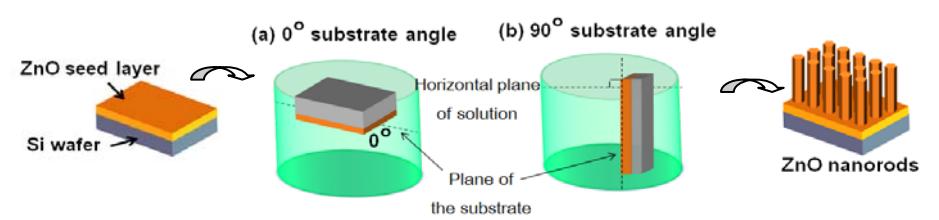
Figure 1 Schematics of the sample preparation for (a) 0° substrate angle and (b) 90° substrate angle ZnO nanorod growth in aqueous solution.
Finally, field emission scanning electron microscopy (FE-SEM, Hitachi) was used to study the microstructure of the ZnO nanorods. Additionally, the crystallinity of ZnO was analyzed by grazing incidence X-ray diffraction (GIXRD, Rigaku). Using a voltage of 40kV and a current of 30 mA, a 20 scanning range from 20° to 70° was explored with a step-size of 0.02.
3 Results and Discussions
Figure 2 shows the SEM images of the morphology of the ZnO nanorods, with insets showing cross-sections of the 3 ratios for Zn(NO<sub>3</sub>)<sub>2</sub>.6H<sub>2</sub>O:HMTA at 0° and 90° substrate angle, respectively. Table 1 shows the diameters, lengths and aspect ratios of the nanorods for these precursor compositions. The lengths of the ZnO nanorods specified in the table do not include the needle part that grows on top of the ZnO nanorods as shown in Figures 2 (a) and (d). We found that at a Zn(NO<sub>3</sub>)<sub>2</sub>.6H<sub>2</sub>O:HMTA ratio of 1:0.5, ZnO needles grew on top of the nanorods such that we can call them nanocandles. One reason for this result is that HMTA generated OH which bonded with Zn<sup>2</sup>, thus forming the ZnO nanorods. The ZnO nanorods' size decreased with growth time due to the depletion of HMTA resulting in discontinuous ZnO growth in the lateral plane. When the Zn(NO<sub>3</sub>)<sub>2</sub>.6H<sub>2</sub>O:HMTA ratio was 1:5, the constant diameters sizes and different lengths were as shown in Figures 2(c) and 2(f). In this case, the excess OH- dissolved part of the ZnO nanorods back into the solution, so that the ZnO nanorods had a limited diameter size. The ZnO nanorods grew in the longitudinal direction instead.
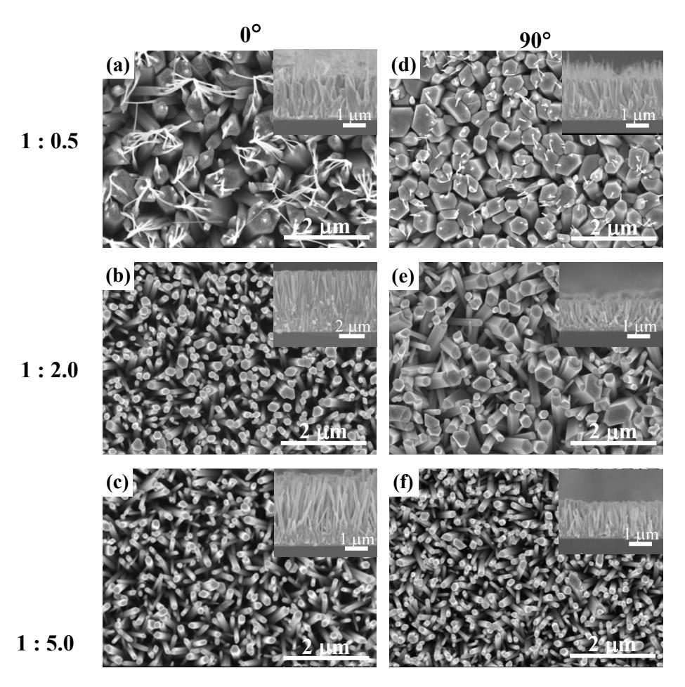
Figure 2 SEM images of ZnO nanorods of six samples, where 2(a) to 2(c) show the 0 substrate angle at 1:0.5, 1:2.0, 1:5.0 ratios of Zn(NO3)2.6H2O: HMTA, and 2(d) to 2(f) show the 90 substrate angle at 1:0.5, 1:2.0, 1:5.0 ratios of Zn(NO3)2.6H2O:HMTA (the insets are corresponding cross-section images).
Furthermore we observed that at a 1:2 concentration ratio of Zn(NO3)2.6H2O:HMTA, the different substrate angles showed a contrast in diameter size. The reason is that when the substrate angle was 90, the ZnO saturated in the solution fell onto the substrate by gravitational force and formed on the lateral planes faster than the longitudinal ones. Hence, the diameter size of the 90 condition increased, as can be seen in Figure 2(e). Meanwhile, ZnO growth at 0 used only thermal energy, without gravitational force, so the longitudinal plane grew faster than the lateral one, as can be seen in Figure 2(b). The aspect ratio of ZnO growth for this condition in Table 1 indicates that the structure of the produced ZnO is nanorods with a highest value of 27.69.
| Table 1 | Relation between diameter size, length and aspect ratio for various | ||||
|---|---|---|---|---|---|
| precursor composition ratios. |
| at 0o | at 90o | |||||
|---|---|---|---|---|---|---|
| Composition Ratio | Diameter (avg) (nm) | Length (m) | Aspect ratio | Diameter (avg) (nm) | Length (m) | Aspect ratio |
| 1:0.5 | 645 | 1.79 | 2.78 | 452 | 1.76 | 3.89 |
| 1:2.0 | 185 | 5.12 | 27.69 | 406 | 1.27 | 3.11 |
| 1:5.0 | 165 | 2.96 | 17.96 | 162 | 1.54 | 9.52 |
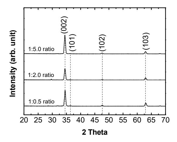
Figure 3 X-ray diffraction pattern of ZnO nanorods at 0 substrate angle for three Zn(NO3)2.6H2O:HMTA ratios.
The result of the crystal structure of the ZnO at 0 substrate angle for three ratios of Zn(NO3)2.6H2O:HMTA were investigated and analyzed from the XRD pattern in Figure 3. The 2 peaks were observed at 34.4°, 47.5° and 62.8°. All the obtained XRD diffraction patterns are according to JCPDS card number 036-1451. The peaks in the XRD spectrum could be assigned to the (002), (102) and (103) crystal planes of the hexagonal wurtzite crystal structure. The crystal structures of ZnO at 0 substrate angle were the same as at 90 substrate angle.
4 Conclusions
The experimental results revealed that the growth of ZnO nanorods can be synthesized using the aqueous solution method. The influence of various HMTA precursor compositions and substrate angles on the morphology of ZnO nanorods growth were studied. It can be concluded that these two preparation parameters, i.e. precursor composition and substrate angle, are correlated to the growth behavior of ZnO structures. We found that the HMTA concentration affects the ZnO chemical reaction. If HMTA is lower than Zn(NO3)2.6H2O, the ZnO cannot grow continuously, while if HMTA is higher than Zn(NO3)2.6H2O, the ZnO can dissolve into the solution and diameter size of ZnO is limited. Moreover, the results indicate that the substrate angle affects the external force that acts on the ZnO particles collected on the substrate. If the ZnO is put at a 90 substrate angle, the lateral growth is faster than the longitudinal growth. Consequently, the optimization of these two preparation parameters is an interesting topic for investigation in a future study.
Acknowledgements
The funding of this work and sputtering deposition was supported by Srinakharinwirot University and Optical Thin-Film Laboratory (OTL), National Electronics and Computer Technology Center (NECTEC), respectively, for which the authors would like to express their sincere appreciation.