
1 Introduction
Since 1981, several reports have been published concerning overexpression of the epidermal growth factor receptor (EGFR) on cancer cells [1-3]. One third of all epithelial cancers, such as head and neck cancer, colorectal carcinoma, nonsmall cell lung cancer (NSCLC) and pancreatic cancer, have been reported to express a high level of EGFR. Overexpression of EGFR indicates tumor malignancy and poor prognosis of a cancer, and also correlates with cell
proliferation, metastases and radiation resistance [4,5]. Therefore, EGFR has been proposed as a target for cancer targeted therapy [2,6].
Nimotuzumab is a humanized monoclonal antibody that binds with domain III of the extracellular region of the EGFR [7]. Nimotuzumab has been used for the treatment of various carcinomas such a glioma, head and neck, lung, renal, colorectal, bladder, prostate, pancreatic, ovarian, cervical and breast cancer. Preclinical and clinical trials performed by Crombet, et al., showed that nimotuzumab has an antiproliferative, proapoptosis and antiangiogenic effect [6,8]. In a randomized open-label, phase-IIb, 5-year study on Indian patients, nimotuzumab was shown to provide a long-term survival benefit to patients with inoperable advanced squamous cell carcinoma of the head and neck [9]. Nimotuzumab has several advantages when compared with other anti EGFRs. These include having a more prolonged half-life in order to give optimum therapy and having no side effects such as skin rash, diarrhea or anaphylaxis, which are common in the use of other anti-EGFRs [3,6,4]. The use of nimotuzumab along with radiation therapy has been shown to enhance the effects of radiation therapy [7]. However, high molecular weight intact nimotuzumab has a poor pharmacokinetic profile [10].
Monoclonal antibody (mAb) fragments are widely used to improve pharmacokinetic profiles [7,8]. MAb fragments have been reported to have better tumor penetration, more rapid blood clearance, lower retention time in non-target tissues and less immunogenicity. Uptake by mAb fragments is eight time faster than by intact monoclonal antibodies, so imaging can be performed on the same day as the administration of radioimmunoconjugate [11].
In recent days, polymeric biomaterials have become the central component of most new drug delivery systems. One of them is a family of water-soluble cascade polymers named poly(amidoamine) (PAMAM) dendrimers. PAMAM dendrimers have a high number of arms and surface amine groups so that they can be utilized to immobilize drugs, enzymes, antibodies or other bioactive agents. PAMAM dendrimers can also be used to improve the amount of therapeutic radionuclides delivered to cancer cells [12,13].
In this study, nimotuzumab-F(ab')2 was used as one of the components of a building block for a new therapeutic radioimmunoconjugate. Lutetium-177 (Lu-177) used as a therapeutic radionuclide with a bifunctional chelating agent of 1,4,7,10-tetraazacyclododecane -1,4,7,10-tetraacetic acid mono-Nhydroxysuccinimide ester (DOTA-NHS-ester). It should be noted that if a high number of DOTA directly binds to nimotuzumab-F(ab')2, it could reduce the ability of the nimotuzumab-F(ab')2 to bind to its receptor. Therefore, in this research PAMAM dendrimer was used for binding a large number of DOTA to produce (DOTA)n-PAMAM, followed by its conjugation onto nimotuzumab-F(ab')2. This technique is expected to offer a large amount of DOTA indirectly bound to nimotuzumab-F(ab')2, which simultaneously binds the Lu-177 without compromising its immunoreactivity.
2 Methods
2.1 Chemicals
Nimotuzumab/TheraCIM was purchased from Kalbe Farma Tbk. Pepsin from porcine gastric mucosa was purchased from Sigma and used without further purification. Monobasic sodium phosphate, dibasic sodium phosphate and N,Ndimethylformamide were purchased from Sigma. Laemli sample buffer, 10x tris/glycine/SDS buffer, Coomasie brilliant blue G-250, Chelex 100 resin, protein standard and protein dye were purchased from Bio-Rad. Acetic acid glacial 100% anhydrous, natrium acetate trihydrate, methanol, natrium hydroxide, hydrochloric acid 32%, sodium chloride, ammonium acetate and ethylene diamine tetraacetic acid were acquired from Merck. Enriched lutetium oxide (60.60%) was purchased from Isoflex. Dendrimer PAMAM G.3 was purchased from Sigma-Aldrich. DOTA-NHS-ester was purchased from Macrocyclics. Traut's reagent (2-Iminothiolane HCl), sulfo-SMCC [sulfosuccinimidyl-4-(N-maleimidomethyl) cyclohexane 1-carboxylate] and dialysis cassettes (20kDa MWCO) were purchased from Thermo Scientific. A PD-10 column was purchased from GE Healthcare. Instant thin-layer chromatography (ITLC SG) was purchased from Gelman Science.
2.2 Instruments
Equipment used for the experiment: thermomixer (Eppendorf), centrifuge (Beckman), mini protean tetra system (Bio-Rad), water purification system (Sartorius Stedim Biotech), high-performance liquid chromatography (Waters) equipped with a size exclusion column (BioSuite) and UV-visible detector (Waters), microplate reader (Biotek Instruments), gamma counter (Nucleus) and dose calibrator (Capintec).
2.3 Preparation and Characterization of Nimotuzumab F(ab')2 Fragments
Nimotuzumab was fragmented using pepsin with a similar method as reported by Hao Hong, et al., [11]. Termination of the fragmentation process, unlike reported by R.D. Haryuni, et al., [14], where Tris HCL 10 mM pH 8 was used, in this project was performed by adjusting the pH of the solution to 8 using 0.1 N NaOH. Samples were analyzed by sodium dodecyl sulfate-polyacrylamide
gel electrophoresis (SDS-PAGE) under non-reducing procedure to identify the optimal digestion condition. Purification of the nimotuzumab-F(ab')2 fragments was carried out using the PD-10 column with phosphate buffered saline (PBS) as the mobile phase. The eluted molecules were detected using Biorad-dye, giving a blue color. The purity of the nimotuzumab-F(ab')2 fragments was evaluated using size exclusion column chromathography with 0.01 N phosphate buffered saline (PBS) pH 7,4 as the mobile phase. Finally, the concentration of the nimotuzumab-F(ab')2 fragments was determined by microplate reader using UV-absorbance at 280 nm.
2.4 Conjugation of DOTA PAMAM Nimotuzumab-F(ab')2 Fragments
Dendrimer PAMAM was added to the sulfo-NHS-DOTA, which had been previously dissolved in 0.1 M phosphate buffer pH 7.2 (mol ratio dendrimer PAMAM to sulfo-NHS-DOTA was 1:96). The pH was then adjusted to 7.2. After 24 hours incubating at 4 °C, the mixture was purified using the PD-10 column with 0.05 M phosphate buffer pH 7.4 as mobile phase. Fractions containing DOTA-PAMAM were collected, followed by addition of 2 iminothiolane-HCl solution (in 0.05 M phosphate buffer pH 7.4, 1 mg/mL) at proportional mol ratio. The mixture was incubated for an hour (in nitrogen gas flow) and then purified using the PD-10 column. The thiolated DOTA-PAMAM fractions were mixed with the maleimide-activated nimotuzumab-F(ab')2 fragments [15]. The maleimide-activated Nimotuzumab-F(ab')2 fragments were prepared by adding sulfo-SMCC (sulfo-SMCC was dissolved in DMF and then diluted with 0.1 M phosphate buffer pH 7.2 to get a concentration of 1 mg/mL) to the nimotuzumab-F(ab')2 fragments, with the mol ratio of sulfo-SMCC to nimotuzumab-F(ab')2 fragments at 10-fold molar excess. The mixture was incubated for 30 minutes at room temperature and then purified using the PD-10 column. The mixture of thiolated DOTA-PAMAM and the maleimide-activated nimotuzumab-F(ab')2 fragments (mol ratio 10:1) was incubated for 24 hours at 4 °C followed by dialysis against 0.25 ammonium acetate pH 7.5 and Chelex 100 with 4 buffer changes every 12 hours.
2.5 Labeling of (DOTA)n-PAMAM-[Nimotuzumab-F(ab')2 Fragments]
177LuCl3 was prepared by iradiating 176Lu2O3 (60.60% enriched) in a Multi Purpose Research Reactor (BATAN, Indonesia) for 4 days. The irradiated target was then moved to a beaker glass and added with 2 mL of H2O2. The mixture was heated until dried and then redissolved in 3 mL of HCl 0.5 N.
The 177LuCl3 (19 mCi) was diluted with 0.25 M ammonium acetate pH 7.5 (1:1 v/v) and then added to (DOTA)n-PAMAM-[nimotuzumab-F(ab')2 fragments]. The pH of the mixture solution was adjusted to 5.5, followed by incubating for 1 hour at 37 °C. Purification of the radioimmunoconjugate, (177Lu-DOTA)n-PAMAM-[nimotuzumab-F(ab')2], was performed using the PD-10 column with 0.01 N phosphate buffer saline pH 7.4 as the mobile phase.
3 Results and Discussion
In a previous study, radioimmunoconjugate 177Lu-DOTA-nimotuzumab with high radiochemical purity was successfully prepared [16]. However, intact monoclonal antibodies tend to have a relatively poor pharmacokinetic profile and tumor penetration, and potential for eliciting host antibody responses [17]. Therefore, in this study we prepared an antibody fragment-based radioimmunoconjugate (177Lu-DOTA)n-PAMAM-[nimotuzumab-F(ab')2] to achieve faster tumor penetration and blood clearance. F(ab')2 fragments were prepared by digestion of whole or intact nimotuzumab using pepsin. The choice for F(ab')2 fragments was due to their bivalent form. These fragments still have two antibody arms that are able to bind to two targets simultaneously, which is important for maintaining prolonged drug residence in tumors and is an important feature for inhibiting tumor growth [18]. Optimization of digestion time at 37 °C (pH 3.5) and a pepsin to nimotuzumab mol ratio of 1:50 for generating nimotuzumab-F(ab')2 without the presence of intact nimotuzumab was achieved at 6 hours of digestion time (Figure 1). SDS-PAGE gel (Figure 1) showed that there were two bands of interest in determining the results of nimotuzumab digestion. The band at 150 kDa indicates intact nimotuzumab while the 100 kDa band shows nimotuzumab-F(ab')2.
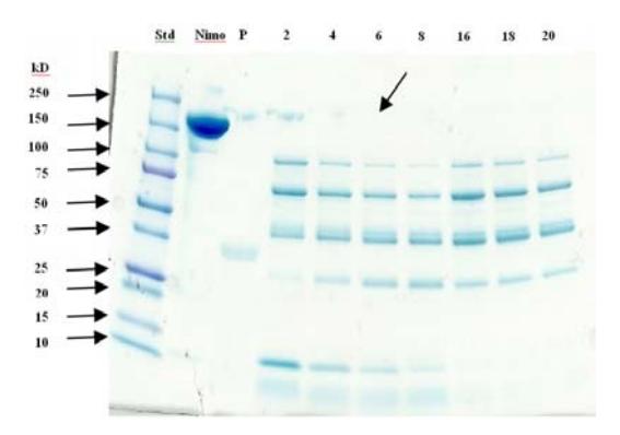
Figure 1 SDS-PAGE of protein standard, intact nimotuzumab, pepsin, and nimotuzumab fragments. Arrows indicate the absent of intact nimotuzumab (150 kD MW) at 6 hours digestion time.
The effect of the pepsin to nimotuzumab mol ratio was investigated by varying it at 1:200, 1:100, 1:50, 1:20, 1:10, 1:5 and 1:1. The nimotuzumab was found completely digested to its F(ab')2 fragments by using pepsin to nimotuzumab with a mol ratio of 1:100 and up to 1:1 (data not shown). When the mol ratio of pepsin to nimotuzumab was 1:20 and up to 1:1 the mixture became turbid, which is allegedly caused by nimotuzumab damage. At this mol ratio, the concentration of pepsin to antibody species is significantly high; the enzyme not only digests the antibody into dimeric F(ab')2 and Fc fragments, but it may also attack the antigen binding site on the Fab. Pepsin is usually used to fragment and cleave proteins on many points to give small peptides (3-30 residues). The active conformation of pepsin appears at an optimal pH of around 2, while at pH above 7, pepsin is in denatured conformation. This denaturation is not fully reversible [19].
Optimization of digestion time and the amount of pepsin are crucial because the affinity of nimotuzumab-F(ab')2 can be diminished or eliminated if the incubation is too long or the concentration of pepsin is too high. The digestion process has been reported to be highly variable, which results in many different protocols for individual antibody species and amounts digested [20]. In further digestion, performed with an incubating time of 6 hours and a pepsin to nimotuzumab mol ratio of 1:100. Termination of the fragmentation process was carried out by adjusting the pH to 8. The generated nimotuzumab-F(ab')2 was then purified from byproducts such as nimotuzumab-Fc, nimotuzumab-Fab, pepsin, etc. using the PD-10 column. The purity of nimotuzumab-F(ab')2 was evaluated using a size exclusion column chromatographic system. Figure 2 shows the chromatograms of nimotuzumab-F(ab')2 before and after purification.
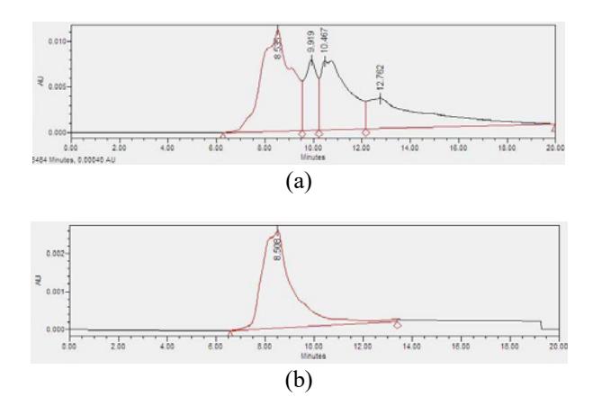
Figure 2 Chromatograms of nimotuzumab-F(ab')2, before (a) and after (b) purification.
It can be seen that before purification there were some peaks that depict the \(F(ab')_2\), Fab, Fc and several small fragments of nimotuzumab (Figure 2a). After purification however, the obtained –nimotuzumab \(F(ab')_2\) was found as a single peak, which indicates high purity (Figure 2b).
The maleimide-activated nimotuzumab-F(ab')<sub>2</sub> was then conjugated to (DOTA)<sub>n</sub>-PAMAM dendrimer, which had been activated previously with Traut's reagent to form (DOTA)<sub>n</sub>-PAMAM-[nimotuzumab-F(ab')<sub>2</sub>]. The main purpose of using PAMAM dendrimer is to increase the number of DOTA indirectly bound to nimotuzumab-F(ab')<sub>2</sub>, which in turn would maximize the number of <sup>177</sup>Lu bounds in the building blocks of (<sup>177</sup>Lu-DOTA)<sub>n</sub>-PAMAM-[nimotuzumab-F(ab')<sub>2</sub>] (Figure 3). As a large amount of <sup>177</sup>Lu-DOTA does not directly bind to nimotuzumab-F(ab')<sub>2</sub>, which functions as a delivery agent of the (<sup>177</sup>Lu-DOTA)<sub>n</sub>-PAMAM-[nimotuzumab-F(ab')<sub>2</sub>] to the EGF receptor, the immunoreactivity of nimotuzumab-F(ab')<sub>2</sub> would be expected to be relatively intact.
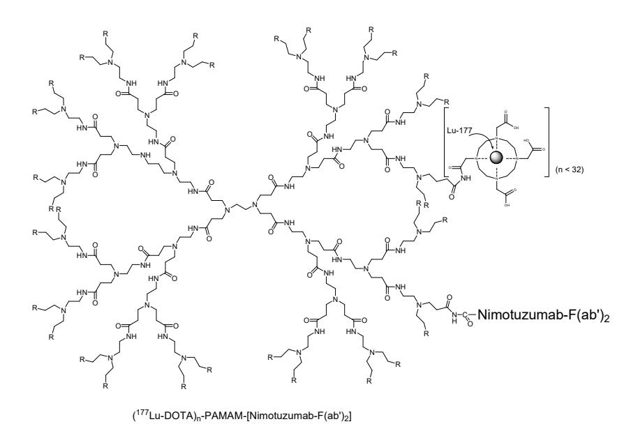
Figure 3 Building block of (<sup>177</sup>Lu-DOTA)<sub>n</sub>-PAMAM-[nimotuzumab-F(ab')<sub>2</sub>]. The figure is created using ChemDraw Ultra 8.0.
Figure 4 shows radiochromatograms of \((DOTA)_n\)-PAMAM-[nimotuzumab-\(F(ab')_2\)] triplicate labeling with \(^{177}Lu\). It can be seen that the radiochemical purity of the \((^{177}Lu\)-DOTA)<sub>n</sub>-PAMAM-[nimotuzumab-\(F(ab')_2\)] resulted from the radiolabeling processes was \(69.59\pm1.66\%\). In order to obtain a radioimmunoconjugate that has radiochemical purity > 95% (requirement for a
(177Lu-DOTA)<sub>n</sub>-PAMAM-[nimotuzumabradiopharmaceutical), the F(ab')<sub>2</sub>] was purified using the PD-10 column. Fractions (250 mL/ fraction) were then retrieved. Each fraction was then measured for its radioactivity by using a dose calibrator. A radiochromatogram of the purified (177Lu-DOTA)<sub>n</sub>-PAMAM-[nimotuzumab-F(ab')<sub>2</sub>] is shown in Figure 5.
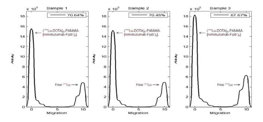
Figure 4 Labeling efficiency of (DOTA)<sub>n</sub>-PAMAM-[nimotuzumab-F(ab')<sub>2</sub>] with <sup>177</sup>Lu radionuclide.

purified (177Lu-DOTA)<sub>n</sub>-PAMAM-Figure 5 Radiochromatogram of [nimotuzumab-F(ab')<sub>2</sub>].
It can be seen that there are two peaks in the radiochromatogram of the purified \((^{177}Lu\text{-DOTA})_n\)-PAMAM-[nimotuzumab-F(ab')<sub>2</sub>]. These peaks represent \((^{177}Lu\text{-DOTA})_n\)-PAMAM-[nimotuzumab-F(ab')<sub>2</sub>] (fraction 11 – 17) and free 1777Lu (1777Lu which does not bind to (DOTA)<sub>n</sub>-PAMAM-[nimotuzumab-F(ab')<sub>2</sub>]). The radiochemical purity of fractions under the (<sup>177</sup>Lu-DOTA)<sub>n</sub>-PAMAM-[nimotuzumab-F(ab')<sub>2</sub>] peak was then analysed using thin-layer
chromatography, where ITLC-SG was used as stationary phase and saline solution as mobile phase. The radiochemical purity of fraction 12-15 was found to be 99.61±0.27% (Figure 6). These fractions were pooled and used for further studies.
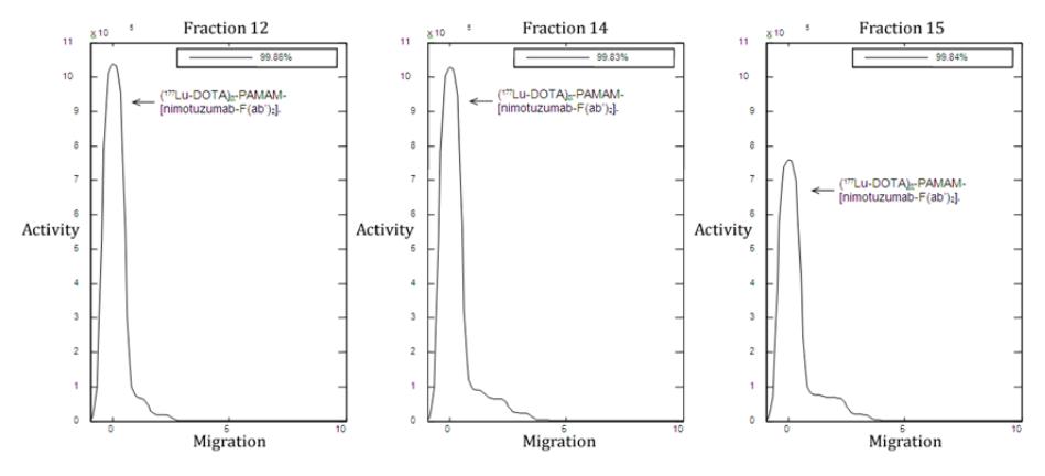
Figure 6 Radiochemical purity of 177Lu-DOTA-PAMAM-[nimotuzumab-F(ab')2].
4 Conclusion
Optimal conditions for digestion of nimotuzumab into F(ab')2 fragments using pepsin were achieved. Nimotuzumab was completely digested at a pepsin to nimotuzumab mol ratio of 1:100, a pH of 3.5, and 6 hours digestion time. Preparation of radioimmunoconjugate (177Lu-DOTA)n-PAMAM- [nimotuzumab-F(ab')2] was successfully conducted, with a labeling yield of 69.59±1.66% and radiochemical purity of > 99% after purification with a PD-10 column. For further research it is advised to perform a biodistribution test of radioimmunoconjugate (177Lu-DOTA)n-PAMAM-[nimotuzumab-F(ab')2] and then compare its pharmacokinetic profile with that of radioimmunoconjugate ( 177Lu-DOTA)n-PAMAM-nimotuzumab.
Acknowledgements
The authors would like to thank Mr. Hambali, Mr. Abidin, Mr. Muhamad Subur and Ms. Sri Setiyowati for preparing the radionuclide Lutetium-177.