
1 Introduction
Black rice is becoming popular for the development of functional foods. Indonesia has many variants of cultivated black rice. One of them is called Cempo Ireng and is grown in several regions of Daerah Istimewa Yogyakarta (DIY), Indonesia. Previous studies have reported that this black rice cultivar has a higher content of anthocyanin (428.38 mg/100g) than other cultivars [1]. Phytochemical studies suggest that black rice bran contains many kinds of secondary metabolites, such as a variety of flavones, tannin, phenolics, sterols, tocols, γ-oryzanols, amino acids, and essential oils. The most abundant functional component of black rice is anthocyanin. Several anthocyanins have been isolated and identified from pigmented rice, including cyanidin 3glucoside, cyanidin 3-galactoside, cyanidin 3-rutinoside, cyanidin 3,5-diglucoside, malvidin 3-galactoside, peonidin 3-glucoside, and pelargonidin 3,5-diglucoside [2].
Breast cancer is one of the most frequent cancer cases for women in the world [3], including in Indonesia. Several studies have focused on cancer prevention and how to reduce this problem. Many researchers used the continuous cell line T47D for their breast cancer cell model. These cells have specific characteristics, such as luminal A subtype, ER<sup>+</sup>, PR<sup>+</sup> and HER2<sup>-</sup> [4,5]. Previous studies have shown that T47D cells express defective p53 protein [6], which regulates apoptosis and the cell cycle, among others [7]. Meanwhile, T47D cells express caspase 3 and 7 wild types. Previous studies suggest that T47D cells have many changes in chromosome number and structure, such as deletion, translocation, and duplication [5].
Some studies revealed that the bioactive phytochemicals from rice bran influence the growth of several kinds of cancer cells through proliferation inhibition, apoptosis induction, and altering the cell cycle [8]. Many kinds of black rice products and extracts demonstrate cytotoxic activities, such as methanolic extract of brewers' rice Temukut on HT-29, Caov3 and HepG2 with an IC<sub>50</sub> of 54.00, 62.33, and 56.00 μg/mL, respectively [9]. In China, anthocyanin-rich extract from black rice showed activity of decreasing MCF-7 breast cancer cell viability and inducing apoptosis of MDA-MB-435 [10]. So far, anthocyanin extract from pigmented black rice of cultivar Heugjinjubyeo (Japan) has demonstrated a cytotoxic effect on human monocyte leukemia cells and altered the G2/M phase of the cell cycle [11]. A recent study revealed that active fractions F<sub>2</sub> and F<sub>4</sub> of methanolic extract of BRB cv Cempo Ireng contain peonidin 3-glucoside and cyanidin 3-glucoside, respectively. However, both compounds were not detected in F<sub>5</sub>. Furthermore, the F<sub>2</sub> and F<sub>4</sub> fractions demonstrated activity of apoptosis induction in HeLa cervical cancer cells [12].
Here, we evaluated the cytotoxicity of methanolic extract fractions of black rice bran (Oryza sativa L cv Cempo Ireng) in T47D cells. Further, we also analyzed the effects of each fraction on the apoptotic and cell cycle alteration responses in T47D cells. The results of this study may support the development of black rice bran for functional food products in Indonesia.
2 Materials and Methods
2.1 Rice Materials and Extraction
Black rice bran (BRB) was obtained from local black rice cv Cempo Ireng, which was harvested by Mr. Jamasto, a black-rice farmer from Sayegan, Sleman
(DIY) in 2015. The rice bran was powdered, passed through a 60-mesh sieve with water content less than 11% and stored in an airtight container. Black rice bran extraction was conducted using the method from Kim, et al. in [13] with some modification. One hundred grams of BRB were macerated using 1 L of methanol–HCL 1 N (85:15) for 48 hours while stirring occasionally to prevent saturation. After being rested for 2 nights, the rice bran mass was filtered using a Whatman No. 1 filter. Solute partitioning was done using 750 ml of n-hexane (Merck) to remove lipid components, followed by maceration, with a 24-hour resting period between each process. Finally, the methanol extract was evaporated using a water bath at 60 °C to get dry extract.
Black rice bran was also extracted with water as a solvent using the procedure from Tan, et al. in [9] with some modification. The BRB (100 g) was macerated with 1 L of distilled water and stirred using a magnetic stirrer at 40 °C for 2 hours, after which the rice bran mass was filtered using a Whatman No. 1 filter. Re-extraction was done twice using 500 mL of water and then the result was evaporated at 60 ºC using a water bath to get dry extract.
The compound groups from either methanolic or water extract were monitored using thin-layer chromatography (TLC) with n-butanol–acetic acid–water (4:1:5) as mobile solute (using the upper part). Silica gel 60 F254 (Merck) was used as the solid phase.
2.2 Cytotoxicity Test using MTT Assay
2.2.1 Cell Line Propagation
Human epithelial breast cancer cells (T47D) were obtained from the culture collection of the Laboratory of Parasitology, Faculty of Medicine Universitas Gadjah Mada (UGM). Propagation of T47D cells was conducted by the following procedures established by this laboratory. Human breast cancer T47D cells were cultured in RPMI medium containing 10% FBS, 2% penicillinstreptomycin and 0.5% of fungizone (Gibco). The cells were grown at 37 °C in 5% CO2 atmosphere (CO2 incubator, Hiraeus) up to 70-80% confluence. For cytotoxicity assay, the cells were calculated and divided into a 96-well cell culture plate, each well containing 5 x 10³ cells.
2.2.2 MTT Assay
The dry extracts of black rice bran were dissolved in 5% of media (2.5% DMSO and 97.5% of RPMI) as the stock solution. Methanol BRB extract stock solution was diluted at concentrations of 2000, 1000, 500, 250, and 125 μg/ml, while water BRB extract was diluted at 4000, 2000, 1000, 500, and 250 μg/ml. Each concentration was treated in a micro-plate well containing 10 μl/ml 5x10³ cells with the maximal volume of media. The maximal DMSO concentration in each well was 0.2%. The cells were exposed to methanol and water BRB extract and incubated for 24 hours and 48 hours respectively. After incubation, the cells were prepared for MTT with the modified procedure from Meiyanto, et al. in [14] and Nurhayati, et al. in [15]. For the MTT assay we used [3-(4,5 dimethyl)-2,5-diphenyl tetrazolium bromide] chemical from Gibco. The assay results were analyzed using an ELISA reader at 595 nm wavelength. The cell death percentage was calculated based on the formula [{(A-D)-(B-C)}/(A-D)] x 100%, in which A = control absorbance, B = extract absorbance, C = extract control absorbance, and D = media control absorbance. The value of half maximal inhibitory concentration (IC50) was determined statistically using probit analysis with the SPSS 13 statistic application.
2.3 Fractionation of Methanolic Extract of Black Rice Bran
The methanolic extract of BRB (1 g homogenized in 3 ml methanol) was fractionated and isolated using preparative thin-layer chromatography with silica gel as stationery phase and methanol as mobile phase. The solvents were butanol–acetic acid–water (4:1:5 v/v) after being concentrated for 17 hours. The extract was dotted on a silica plate using a capillary pipe at about 1.5 cm from the bottom edge. The silica plate was put in a solvent jar and incubated for 3 hours (when the eluent reached about 1.5 cm from the upper edge). The bands on the plate were exposed to UV 254 nm and UV 366 nm. The bands were marked on the Rf side and scraped carefully. The fractions were macerated using methanol–HCl 1% to eliminate the silica powder. Each fraction was filtered and dried up using a water bath at 60 °C [12].
2.4 Cytotoxicity Test of Fractions
Each fraction was tested for cytotoxic activity in the T47D cells using the MTT method with dosage > IC50 of methanolic black rice bran extract.
2.5 Apoptosis and Cell Cycle Analysis Using Flow Cytometer
2.5.1 Apoptosis Analysis
Apoptotic cell measurement was performed using a flow cytometer and an Annexin V-FLUOS staining kit. The cell suspension containing 2.5 x 10⁵ cells/well was cultured in a 6-well plate with a final volume of 2000 μl and incubated for 24 hours. The cells (T47D) were treated using 2000μl/well of active fractions of the methanolic BRB extract and doxorubicin for 48 hours. The cells were harvested using 0.25% trypsin and washed with PBS. The cells were treated using 100 μl of the Annexin V-FLUOS staining kit and incubated in dark conditions for 10 minutes in a dark room. The obtained data of apoptotic and necrotic cells were analyzed using FACSCalibur flow cytometer (Becton-Dickinson).
2.5.2 Cell Cycle Analysis
Cell cycle analysis was performed using the flow cytometer method [16]. The suspension containing 2.5 x 10⁵ cells/well was cultured in a 6-well plate with a final volume of 2000 μl. The cells were treated with 2000 μl of active fractions of methanolic BRB extract with half of the IC50 concentration and incubated for 48 hours and subsequently harvested using trypsin-EDTA. Cells were harvested using a centrifuge (2000 rpm, 30 seconds). The cells were mixed with flow cytometer solution (25 µl PI + 1µl RNAase + 0.5 µl Triton-X + 500 µl PBS) and incubated for 10 minutes in a dark room. The cell suspensions were homogenates and transferred into a flow cytometry tube. The flow cytometer data were analyzed using a FACSCaliybur flow cytometer (Becton-Dickinson).
2.6 Statistical Analysis
Quantitative data are expressed as (mean ± S.D.). Statistical differences between control and samples were determined by one-way ANOVA (Kruskal-Wallis analysis) at a limit of p < 0.05 from 3 independent experiments conducted in triplicate. For comparison between two groups, the data were analyzed using the Mann-Whitney U test. The apoptotic cell data were analyzed using Microsoft Excel 2007, while the cell cycle data were analyzed using flowing software.
3 Results and Discussion
3.1 TLC Profiles of Black Rice Bran Extract
Black rice bran was extracted using methanol, water, and n-hexane solvents. Methanol and water are polar solvents that can extract polar compound groups such as phenolics and flavonoids including anthocyanin. In contrast, n-hexane is a nonpolar solvent that can attract nonpolar compound groups such as terpenoids, carotenoids, and fatty acids. The black rice bran yield from water extraction (28.5%) was higher than that of methanol and n-hexane extraction (20.6% and 3.1% respectively).
From the chromatographic profiles of TLC separation of the methanolic extract and water extract of BRB showed 6 and 5 spots respectively with Rf value ranging from 0.0 to 0.97 (Figure 1). UV 254 nm exposure of the TLC plate showed that the dominant spot in the methanolic extract consisted of flavonoids (Figure 1b) with a color and Rf similar to rutin (as flavonoid standard). This result was confirmed by detection using Sitroborat staining under UV 366 nm, which showed yellow fluorescence, indicating flavonoid compounds.
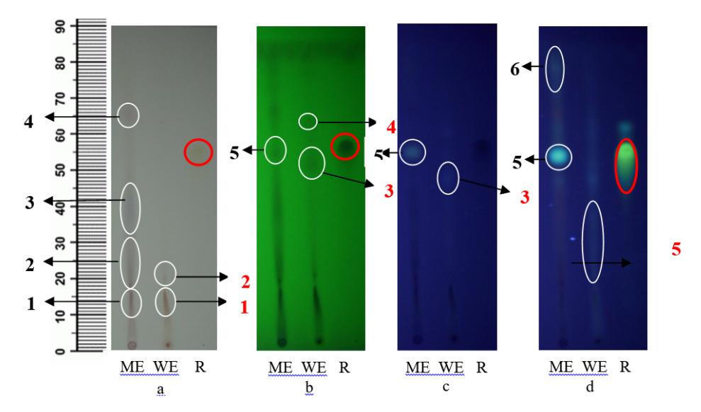
Figure 1 Thin-layer chromatographic profile of methanolic and water extract of black rice bran. Mobile phase of n-butanol–acetic acid–water (4:1:5) detected by a) visible light, b) UV 254 nm, c) UV 366 nm, d) Sitroborat staining and UV 366 nm. ME = methanolic extract; WE = water extract; R = rutin standard. The arrows indicate pointed spots with black numbers for the methanolic extract and red numbers for the water extract. The red cycles indicate pointed rutin spots.
3.2 Cytotoxicity Test of Methanolic Extract Fractions of Black Rice Bran
The cytotoxicity of the methanolic extract of black rice bran on T47D cells can be seen in Table 1 and Figure 2. The IC50 value of the methanolic extract was still under the maximal concentration of tumor growth inhibition baseline according to Meyer, et al. in [17], i.e. 1000 µg/ml. The cytotoxic response shown by the T47D cells due to the methanolic BRB extract indicates compounds that can potentially prevent breast cancer growth. Hui, et al. in [10] reported that ethanolic extract of anthocyanin-rich components of black rice from China [18] showed cytotoxic activity and induced apoptosis in a MCF-7 breast cancer cell line. We used methanolic extract instead of ethanolic extract because methanol has a lower boiling point than ethanol and water. The differences in the primary cells of breast cancer between T47D cells and MCF-7 cells may contribute to different responses to those extracts.
Table 1 IC50 values of black rice bran methanolic extract and water extract on T47D cells (48 h incubation).
| Treatment | IC50 for T47D cells (µg/ml) |
|---|---|
| Methanolic extract | 522.14 ± 24.73 |
| Water extract | 6602.76 ± 792.91 |
| Doxorubicin | 0.064 ± 0.013 |
Although water extract is a polar solvent, its polarity is lower than that of methanol. From the chromatographic profile (Figure 1) the differences between the spots in the methanolic extract and the water extract can be seen. Hence, it can be said that there are different compounds that dissolve in both solvents. The compounds in the methanolic extract of black rice may have synergism with the cytotoxicity activities in the T47D cell.
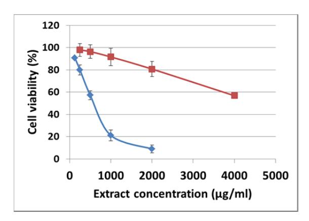
Figure 2 Cell viability of T47D after treatment with methanol (blue line) and water (red line) extracts of black rice bran.
Figure 3 Condition of T47D cell morphology during treatment with methanolic extract of black rice bran: a) T47D cell growth in RPMI media; b) T47D cells after treatment with methanolic extract of BRB; c) T47D condition after 4 hours of treatment with 3-(4,5-dimethyl)-2,5-diphenyl tetrazolium bromide during MTT assay. Cell morphology after treatment is indicated by black arrows. Cell culture multiplication at 400x.
The results of cytotoxic activity of the methanolic extract of BRB on T47D cell morphology are shown in Figure 3. Several T47D cells shrank due to the defect of cytoskeleton, which caused morphological changes. Some cells exhibited loss of adhesion to neighboring cells or the extracellular matrix [19]. After we got the IC50 of the methanolic BRB extract on the T47D cells, we proceeded to fractionate the extract using preparative TLC.
3.3 Cytotoxic Activities of Fractions
The results of the fractionation of the methanolic extract indicated at least 6 fractions with Rf values ranging from 0 to 0.97 (Figure 4). Each fraction was scraped and weighed, and then dissolved and macerated in methanol–HCl 1 N (85:15). Figure 4 shows that fraction F5 was dominant (0.51 g) and had a dark color (black).
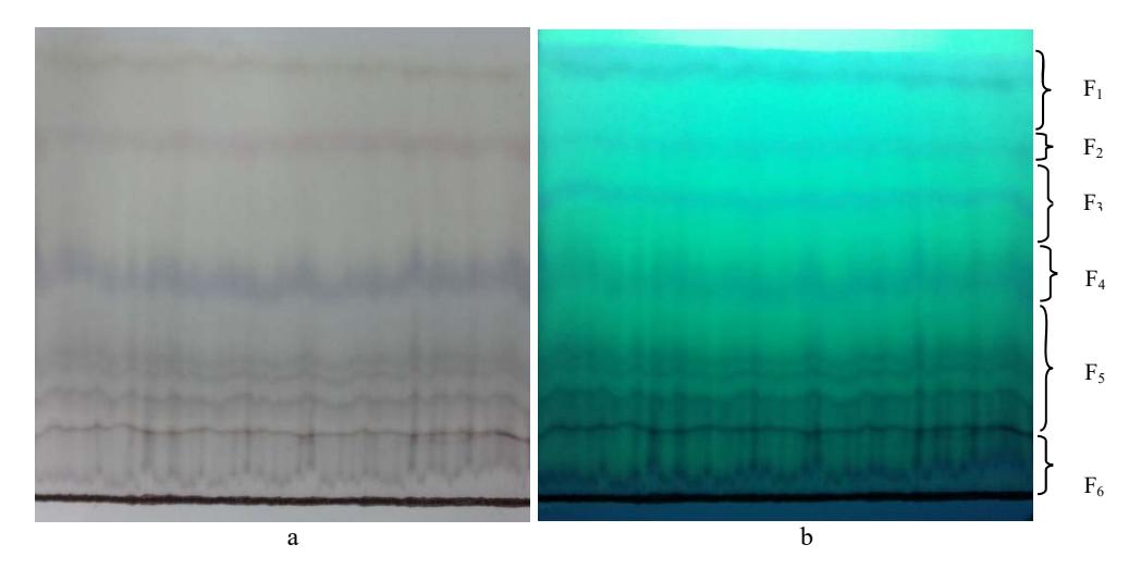
Figure 4 Chromatograph of preparative TLC of methanolic extract of rice bran. Mobile phase = n-butanol–acetic acid: water (4:1:5, upper side). a) Detected by visible light, b) detected by UV 254 nm. Six fractions were scraped from the TLC silica plate. The fraction codes used are: F1, F2, F3, F4, F5 and F6.
The values of cytotoxicity, or inhibition of concentration, at 50% living cells (IC50) from the 6 fractions of the BRB methanolic extract in T47D cells can be seen in Table 2. The results showed that all fractions showed cytotoxic activity with IC50 values lower than 100 μg/ml. The lowest IC50, i.e. the strongest cytotoxic activity, was shown by F3, followed by F2, F5, and F4 with values of 60.17, 64.33, 66.62, 71.22 μg/ml respectively.
Table 2 IC<sub>50</sub> Values of preparative TLC fractions on T47D cells incubated for 48 hours.
| Fraction | IC50 on T47D cells (μg/ml) |
|---|---|
| \(\mathbf{F}_{1}\) | \(90.54 \pm 3.52\) |
| \(F_2\) | \(64.33 \pm 0.61\) |
| \(F_3\) | \(60.17 \pm 1.72\) |
| \(F_4\) | \(71.22 \pm 3.11\) |
| \(F_5\) | \(66.62 \pm 2.53\) |
| \(F_6\) | \(89.01 \pm 2.63\) |
3.4 Apoptotic Induction of Active Fractions
Most of the BRB fractions showed lower IC<sub>50</sub> values than the BRB methanolic extract. However, these cytotoxic activities were still lower compared to those of the drug doxorubicin, which is usually applied as chemotherapy treatment for breast cancer patients. Thus, those 4 fractions were selected for further analysis to find out the cell death mechanism due to either apoptosis or necrosis (Table 3).
Table 3 Percentages of T47D cell deaths caused by preparative TLC fractions of methanolic extract of black rice bran.
| Treatment | Cell percentage (%) | |||
|---|---|---|---|---|
| Live | Apoptotic | Necrosis | ||
| Control cells | \(90.12 \pm 0.67^{e}\) | \(8.39 \pm 0.16^{a}\) | \(1.50 \pm 0.50^{a}\) | |
| Fraction 2 | \(5.83 \pm 0.42^{a}\) | \(75.39 \pm 0.43^{e}\) | \(18.79 \pm 0.85^{c}\) | |
| Fraction 3 | \(7.04 \pm 0.14^{bc}\) | \(58.78 \pm 0.71^{c}\) | \(34.19 \pm 0.86^{e}\) | |
| Fraction 4 | \(7.6 \pm 0.24^{c}\) | \(68.87 \pm 1.82^{d}\) | \(23.53 \pm 1.58^{d}\) | |
| Fraction 5 | \(6.51 \pm 0.38^{ab}\) | \(60.36 \pm 2.07^{c}\) | \(33.14 \pm 1.68^{e}\) | |
| Doxorubicin | \(51.77 \pm 0.21^{d}\) | \[41.30 \pm 0.08^b\] | \(6.94 \pm 0.28^{b}\) | |
The results showed that all fractions (F<sub>2</sub>, F<sub>3</sub>, F<sub>4</sub>, and F<sub>5</sub>) could induce more cell apoptosis than doxorubicin, which only inhibited about 41.30%. Meanwhile, the active fraction with the highest apoptotic induction was F<sub>2</sub> (75.39%) followed by F<sub>4</sub> (68.87%). The F<sub>2</sub> and F<sub>4</sub> contain peonidin 3-glucoside (0.012 μg/ml) and cyanidine 3-glucoside (0.14 μg/ml), as reported by Pratiwi, et al. in [12]. This result is confirmed by a result from Hui, et al. in [10], which suggests that anticancer compounds from rice bran could induce more apoptosis than necrosis in cancer cells. A previous study has shown that cyanidin 3-glucoside and peonidin 3-glucoside are potential candidates for cancer chemoprevention. These compounds can reduce cell growth via apoptosis induction in human breast carcinoma HS578T cells [20]. So far, it can be said that T47D cells are more responsive to BRB extract fractions than HeLa cells, as has been reported recently [12]. On the other hand, the T47D cells were less responsive to doxorubicin compared to the extract fraction treatment (Table 3). Sugimoto, et al. in [21] has suggested that necrosis in cancer cells caused by doxorubicin is due to the ability of the cells to reduce doxorubicin compounds so they become semiquinone free radicals, which causes reactive oxygen species (ROS) induction. In this situation, doxorubicin can destroy DNA or the cell membrane. However, in this study, the percentages of apoptotic cells of T47D treated by BRB extract fractions (F2, F3, F4, and F5) were higher than those of cells with doxorubicin treatment. This could be due to enhancement of the doxorubicin resistance effect on T47D cells [13].
3.5 Active Fractions Effect on T47D Cell Cycle
The distribution of cell cycle G0/G1, S, and G2/M-phases in the T47D cells was analyzed using a flow cytometer. The result can be seen in Figure 5. This result demonstrates that the active fraction of F5 had the highest inhibition (19.25 ± 0.08%) on the T47D cell cycle at S-phase arrest. The compounds of fraction F5 of the methanolic extract of black rice bran significantly increased the S-phase arrest percentage. This inhibition may prevent overt DNA damage [19]. However, Hyun and Chung [11] have reported that anthocyanin cyanidin and malvidin from pigmented black rice extracts (O. sativa cv. Heugjinjubyeo) show cytotoxic activity on human monocyte leukemia cells and indeed induce G2/M-phase arrest. Schantz, et al. in [22] suggests that anthocyanin from bilberry fruits has antioxidant activity by decreasing ROS numbers and total glutton (tGSH) in HT-29 and Caco2 cells. However, the result from the present study still leaves open several possible mechanisms of inducing S-phase arrest in T47D cells. This question needs to be clarified.
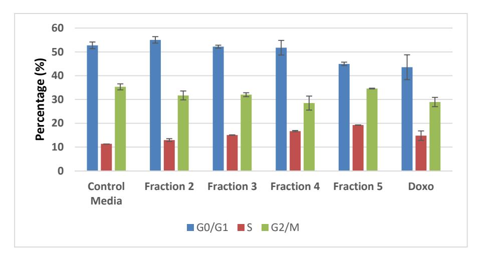
Figure 5 Percentage of T47D cell deaths after treatment with preparative TLC fractions. Fraction 5 showed the highest percentage of S-phase arrest compared to the other fractions.
4 Conclusions
Methanolic extract of black rice bran (cv Cempo Ireng) can prevent the growth of breast cancer T47D cells. Fraction F3 had the lowest IC50 on T47D cells. Fractions F2 and F4 induced apoptosis in T47D cells better than the other fractions and the cancer drug doxorubicin. However, fraction F5 had the highest percentage of T47D cell cycle arrest in S-phase compared to the other fractions, media control, and treatment with doxorubicin.
Acknowledgements
We would like to thank the Ministry of Research, Technology and Higher Education, Republic Indonesia for the research funding under the National Research Competitive Projects 2015-2016.