
1 Introduction
Anthocyanins are natural pigments derived from plants or fruits as secondary metabolites. The pigments have colors that vary between red, violet, and blue. They belong to flavonoid group and have some beneficial effects on human
Received November 6th, 2017, Revised April 17th, 2018, Accepted for publication May 25th, 2018. Copyright © 2019 Published by ITB Journal Publisher, ISSN: 2337-5760, DOI: 10.5614/j.math.fund.sci.2019.51.1.3
health, such as antioxidant, anti-inflammation, chemoprevention activity [1]. Some studies reported that antioxidant activity of anthocyanins is greater than that of others like carotenoid, phenol, etc [2]. Anthocyanins are also claimed that they can improve vision, protect from cardiovascular damage, and have anti-diabetic properties [3].
Beyond their activities, anthocyanins have low stability during processing and storage. Their stability is influenced by some factors like temperature, light, pH, oxygen, concentration, and others [4]. Generally, basic structure of anthocyanins is anthocyanidin which form glycosidic bond with sugar group. Anthocyanins structures are different which depend on pH level. They are flavylium cation, quinoidal base, carbinol base, chalcone that arising in pH 1-3, 3-4, 5, 6 respectively in Figure 1 [5].
O+ O Sugar OH OH HO OH HO O OH O Sugar O OH HO O OH O Sugar OH OH HO O Sugar OH OH HO OH OHO Flavylium cation (AH+) (red) pH 1-3 Quinoidal base (A) (violet) pH 3-4 Chalcone (C) (yellow) pH 6 Carbinol base (B) (colorless) pH 5 -H+ -H+ / +H2O tautomerization
Figure 1 Structures of anthocyanin at different pH condition.
Anthocyanins stability will decrease with increasing pH level so the highest stability of anthocyanins is obtained in flavylium cation structure [6]. Temperature affects stability of anthocyanins by shifting anthocyanins structure to carbinol and chalcone with increasing temperature. Higher temperature can
induce hydrolysis of glycosidic bond of anthocyanins and produce opened and unstable anthocyanidin [7]. Oxygen presence has a role in decreasing stability by oxidizing directly or through oxidator enzyme [8]. Light also induces oxidation process by releasing energy which will shift anthocyanins structure to chalcone [9]. Regarding factors lowering anthocyanins stability, anthocyanins microencapsulation has been proposed to overcome them [10].
Purple-fleshed sweet potato (Ipomoea batatas L.) is one of the anthocyaninenriched food stuffs which contains 1400 mg of anthocyanins / 100g of dry weight. Its anthocyanins stability is more than others sources like blueberry, blackberry, strawberry, purple cabbage, because anthocyanins presence is in acylated structures [11]. It has been reported that acylated anthocyanins have higher antioxidant and anti-mutagenic activity. It has been studied that purplefleshed sweet potato anthocyanins have high stability in condition pH of 3-4 [12].
Microencapsulation is a process aimed to protect active substance by covering it so that active substance can be avoided from interference of external factor, such as temperature, pH, and light. Active substance which has role as core material will be protected by carrier material such as polymer. One of microencapsulation technique for protecting anthocyanins is emulsificationcrosslinking which has over strong points. They are high efficiency in production scale and avoidance from high temperature penetration. The weakness of the technique is possibility that emulsion residue cannot be used again.
Chitosan is one of natural polymer that can be used as carrier material in microencapsulation. The polymer derived from chitin has glycosidic bond between N-acetyl glucosamine group and D-glucosamine group [13]. In acidic environment, amine groups will be protonated become polycation so they can interact ionically with polyanion or multivalent anions forming a complex. Moreover, chitosan has biocompatibility properties such as great mucoadhesive and permeability properties in biological surface, and its ability in increasing active substance absorption through intercellular space [14].
Anthocyanins microencapsulation using emulsification-crosslinking technique with chitosan as carrier material becomes selected technology. The solubility of chitosan and anthocyanins in aqueous phase is relatively high so the emulsion system that can be applied is water in oil (W/O) emulsion. It is needed crosslinkers in the process to get stronger droplets of chitosan, one of them is sodium tripolyphosphate (NaTPP) because of its ability to be ionized and becomes polyanion in solution so that it will interact ionically with protonated chitosan. Microencapsulation of anthocyanins using emulsification-crosslinking with chitosan and NaTPP is infrequently conducted, even the parameters during anthocyanin microencapsulation have not studied yet.
In this study, we investigated process conditions during microencapsulation that influenced properties of microcapsules, that were stirring intensity and carrier material (polymer) concentration, pH of chitosan and NaTPP solution, and also ratio of anthocyanins to polymer concentration. Microstructure of microcapsules and interaction among substances were also studied.
2 Material and Methods
2.1 Material
Chitosan was purchased from PT. Biotech Surindo with deacetylation degree > 85% and viscosity 130 cps. Purple-fleshed sweet potatoes (Ipomoe batatas L.) were obtained from market "Pasar Telo", Karangkajen Yogyakarta supplied from Ngawi, East Java. Sodium tripolyphosphate (NaTPP) was technical grade 85% and was purchased from Sigma-Aldrich, US. Sorbitan monooleate (Span 80) was also purchased Sigma-Aldrich, US. Technical grade ethanol 96% and N-hexane was obtained from CV. General Labora, Yogyakarta. Citric acid 99% was purchased from PT. Bratachem. Paraffin liquid was obtained from CV. Alfa Kimia, Yogyakarta. Potassium chloride, sodium acetate trihydrate, and analytical grade ethanol and acetic acid were purchased from Merck, US. 1,1- Diphenyl-2-picrylhydrazyl (DPPH) was obtained from Sigma-Aldrich, US.
2.2 Methods
2.2.1 Preparation of Anthocyanins Extract
Peeled and cleaned purple-fleshed sweet potatoes were sliced across section and were formed to chips with thickness 0.2-0.4 cm. Then, chips were blanched for 5 min and shock cooled. Blanched chips were homogenized and extracted using solvent of 3% (w/v) citric acid in ethanol 96% at extraction temperature 35 °C and dark condition for 4 h with chips to solvent ratio was 100% (w/v). Supernatant was separated from solid residue using double filter cloth and then was resumed using Buchner Funnel with vacuum through Whatman no.41 filter paper. Finally, supernatant was evaporated at temperature 50 °C under vacuum condition with rotary evaporator (BUCHI R-114, Switzerland) in order to get concentrated anthocyanin extract with concentration 70-80 °Brix.
2.2.2 Preparation of Anthocyanin-Loaded Microcapsules
Briefly, anthocyanin-loaded microcapsules were prepared by emulsificationcrosslinking technique within factors influencing microcapsules were varied and optimized, that were stirring intensity and carrier material (polymer) concentration, pH of chitosan and NaTPP solution, and also ratio of anthocyanins to polymer concentration. Generally, microencapsulation process was depicted at Figure 2. Chitosan was dissolved completely in 0.2 M acetate buffer solution with pH varied at 2.8; 3.0; 3.4; 3.8; and 4.2. Then, anthocyanins extract was dissolved and homogenized in chitosan solution. The solution was used as aqueous phase. Aqueous phase was dropped gradually in continuous phase (sorbitan monooleate : paraffin liquid in volume ratio of 2%) with aqueous to continuous volume ratio of 10%, while stirring was carried out to get water in oil (W/O) emulsion by using mechanical agitation with flat 2-blade impeller. Emulsification was conducted for 1 h until stabile emulsion was gained. Afterward, emulsion droplets were solidified by dropping sodium tripolyphosphate (NaTPP) in water which concentration of NaTPP was 7.5% (w/v). The solution was called as cross-linker solution. Ratio of cross-linker to aqueous solution was 200% (v/v). Solidification was carried out for 4 h until getting stronger microcapsules. Obtained microcapsules were collected, then filtered and washed using N-hexane for omitting oil residue. Finally, microcapsules were rinsed by ethanol, stored and dried in vacuum desiccator.
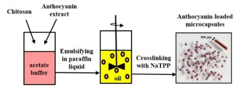
Figure 2 Schematic depiction of anthocyanin microencapsulation using emulsification-crosslinking technique.
2.2.3 The Studying of Microencapsulation Parameters
Investigating of factors influencing microcapsules was conducted step by step (in stages). Firstly, stirring intensity (varied in 1200 and 1600 rpm) and carrier material concentration (varied in 1; 2; and 3% (w/v)) were investigated. The best variables in this stage were used as independent variable in investigating effect of pH of chitosan and NaTPP solution varied in 2.8; 3.0; 3.4; 3.8; 4.2. Finally, the best and selected variable of pH was used as independent variable in evaluating influence of ratio of anthocyanins to polymer concentration varied in 75%; 100%; and 150% (w/w). Parameters of anthocyanin microcapsules used as consideration were efficiency of encapsulation, mean diameter of microcapsules, particle size distribution, and antioxidant activity. The best microcapsules that was result of the optimization of anthocyanins to polymer concentration ratio were investigated by scanning electron microscopy (SEM) and FT-IR analysis for exploring their microstructure and interaction among substances.
2.2.4 Characterization of Microcapsules
2.2.4.1 Encapsulation Efficiency
Encapsulation efficiency indicates effectiveness of carrier material in encapsulating and holding core material. Encapsulation efficiency (EE) was calculated according the Eq. (1) developed by Ye, et al. in [15] :
\[EE(\%) = \frac{\text{encapsulated anthocyanin total content}}{\text{added anthocyanin total in microencapsulation}} x100\%\] (1)
Added anthocyanins total content in microencapsulation refers to anthocyanins total content of anthocyanins extract added in microencapsulation process. Anthocyanins total content both in microcapsules and extract were determined according to pH-differential method [16]. To obtain encapsulated anthocyanins total content, extraction of anthocyanins from microcapsules was conducted initially. About 100 mg of microcapsules was added by 3 ml of HCl-ethanol solvent in which volume ratio of HCl 2M to ethanol 70% was 2/3, mixed using vortex for 5 min, extracted at 4°C and dark condition for 12 h and then followed by centrifugation at 5000 rpm for 10 min at 35 °C to obtain supernatant. It was used two buffer in pH-differential method that were potassium chloride buffer 0.025 M pH 1.0 and sodium acetate buffer 0.4 M pH 4.5. Amount of extract was added to each buffer with volume ratio of 1/6. Then, absorbance of each liquids were measured UV-Vis Spectrophotometer (Dynamica HALO RB-10 Spectrophotometer) at wavelengths of 530 and 700 nm. Total anthocyanins content (TAC) was expressed as cyanindin-3-glucoside according to the following Eq. (2):
\[TAC \binom{mg}{L} = \frac{A \times MW \times DF}{E \times L}\] (2)
where A = (A -A ) - (A -A ) 530 700 pH 1.0 530 700 pH 4.5 ; MW (molecular weight of cyanidin-3-glucoside) = 449.2 g/mole; DF (dilution factor) = 7000, because of conversion from gram to milligram ; E (Molar absorptivity of cyanidin-3 glucoside) =26900 L/mole.cm ; L (cell path length) = 1 cm. Determination of total anthocyanins content of extract was almost similar to that of microcapsules. The difference was anthocyanins extract not need to be extracted. An aliquot of anthocyanins extract was dissolved directly by solvent HCl 2M: ethanol 70% (2/3 v/v). Furthermore, total anthocyanins content of extract solution was also analyzed by pH-differential method.
2.2.4.2 Analysis of Antioxidant Activity
Antioxidant activity of anthocyanins in both microcapsules and extract were determined according DPPH antiradical procedure [17]. In the analyzing of antioxidant activity, DPPH standard curve was previously made by plotting absorbance of DPPH solution measured in Microplate Reader (Thermo Scientific Multiskan GO Type 1510, Thermo Fisher Scientific, Finland) at 515 nm to DPPH concentration (0 – 120 µM). DPPH was dissolved and diluted by using ethanol 80%. Analyzing of antioxidant activity of sample was initiated by extracting about 100 mg of sample with 1.5 ml solvent of acetic acid 0.5% (v/v) in ethanol 40%. Extraction was conducted at 4o C and dark condition for 2 h. Furthermore, supernatant obtained by centrifuging extracted solution was called main solution. The solution was diluted by using acetic acid 0.5% in ethanol 40% with dilution factor of 2; 4; 8; 16; 20; and 40x. While, 75 µM DPPH in ethanol 80% solution was made. Into 96-well microplate, it was filled by the mixing solution with composition as following:
- 1. About 100 µl sample solution (either main sample or diluted main sample) was added to 100 µl DPPH solution 75 µM. The mixed solution was called as sample solution.
- 2. About 100 µl solution of acetic acid 0.5% (v/v) in ethanol 40% was added to 100 µl DPPH solution 75 µM. The mixed solution was called as control solution.
- 3. About 100 µl sample solution (either main sample or diluted main sample) was added to 100 µl ethanol 80%. The mixed solution was called as sample blank solution.
- 4. About 100 µl solution of acetic acid 0.5% (v/v) in ethanol 40% was added to 100 µl ethanol 80%. The mixed solution was called as blank solution.
Filled microplate was entered to Microplate Reader, incubated at 30 °C and dark condition for 30 min. Absorbance of sample, control, sample blank, blank solution were measured at 515 nm. Absorbance of control solution subtracted by absorbance of blank solution was absorbance value used for searching initial DPPH concentration according to obtained standard curve. While absorbance of sample solution subtracted by absorbance of sample blank solution was absorbance value used for searching remaining DPPH concentration. Then, it was calculated % remaining DPPH according Eq. (3) as following:
% remaining DPPH = \[\frac{[remainingDPPH]}{[initialDPPH]} \times 100\%\] (3)
Efficient concentration (EC50) was sample concentration reducing 50% of DPPH. EC50 was obtained by plotting sample concentration to % remaining DPPH and the value was concentration which gave remaining DPPH of 50%. Antioxidant activity was expressed as antiradical power (ARP) for the clarity. ARP was formulated as 1∕EC50. The larger the ARP, the higher antioxidant activity.
2.2.4.3 Size of Microcapsules and Particle Size Distribution
Microcapsules diameter and particle size distribution were determined by using an optical microscopy images taken by Dino-Lite Digital Microscope AM-311, Japan. Image analysis was carried out by Image-Pro software. Mean diameter and particle size distribution were determined according to data of at least 200 microcapsules. Mean diameter was calculated as average of all microcapsules diameter. Particle size distribution obtained by Microsoft Excel was histogram that depicting percentage of amount fraction correlated to microcapsules diameter.
2.2.4.4 Microstructure Characterization
Morphology of microcapsules was investigated by Scanning Electron Microscope (JEOL with JSM-6510 series, Japan). Previously, sample was coated with gold, then scanned with SEM at 10 kV.
2.2.4.5 Fourier Transform Infrared Spectrophotometry (FT-IR) Analysis
For convincing interaction of anthocyanins, chitosan, and NaTPP, FT-IR analysis with Shimadzu IR Prestige-21 was carried out for anthocyanin, chitosan, NaTPP, anthocyanin-loaded microcapsules, and released anthocyaninloaded microcapsules. Amounts 10 mg of sample was prepared into KBr pellet. Furthermore, the sample was transmitted by infra-red spectrum at wavelength of 400–4000 cm-1.
3 Results and Discussion
3.1 Influence of Stirring Intensity and Carrier Material Concentration
Stirring is one of mechanical energy form applied in emulsification for increasing interfacial area so that droplet produced is smaller. Such as stirring, carrier material concentration is also important factor because the substance has role for entrapping bioactive.
According to Table 1 and Figure 3(a), stirring intensity of 1600 rpm produced microcapsules with smaller mean diameter and narrower particle size distribution instead of 1200 rpm at similar carrier material (chitosan) concentration. It was involved with emulsification that higher stirring intensity would produce higher shear rate and energy. The energy was used for breaking droplets became smaller size. Besides that, higher shear stress produced turbulence by which coalescence and aggregation could be prevented. Stirring intensity also influenced encapsulation efficiency. Encapsulation efficiency was higher with increasing stirring intensity. It was involved with emulsification and crosslinking process. Higher stirring intensity would produce smaller emulsion droplets. It meant that the droplets had higher surface area per volume, so that mass transfer flux of cross-linker was higher, further crosslinking process and solidification happened faster by which core material was obstructed for moving outside.
Table 1 The influence of stirring intensity and carrier material concentration on mean diameter and encapsulation efficiency.
| Carrier material concentration (%) | Stirring intensity (rpm) | Mean diameter (µm) | Efficiency of encapsulation (%) |
|---|---|---|---|
| 1 | 1200 | 359.5 | 3.35 |
| 1600 | 328.9 | 7.56 | |
| 2 | 1200 | 384.0 | 9.40 |
| 1600 | 323.1 | 12.41 | |
| 3 | 1200 | 428.4 | 17.10 |
| 1600 | 375.7 | 21.82 |
Table 1 showed that mean diameter of microcapsules was bigger and particle size distribution was broader with increasing of carrier material concentration for similar stirring intensity. Higher concentration of carrier material (chitosan) indicated higher viscosity of system which represented internal friction of fluid. The higher internal friction of fluid, the higher energy needed for fluid moving. Carrier material (chitosan) concentration contributed on efficiency of encapsulation. It can be seen at Table 1 that the higher chitosan concentration used, the higher encapsulation efficiency obtained. Carrier material concentration of 3% (w/v) gave highest encapsulation efficiency for the same of stirring intensity. Higher carrier material concentration induced increasing of system viscosity which obstructed anthocyanins migration from inside to outside of internal phase.
The result may be due to the bonding of chitosan molecule with either crosslinker (NaTPP) or other chitosan molecule. Higher chitosan concentration caused chitosan molecules was closer to each other, so the distance among molecules would be shorter which it increased chitosan – tripolyphosphate
(TPP) intermolecular crosslinking. It was also formed stronger intermolecular hydrogen bonding by higher chitosan concentration. Those made tendency that more chitosan molecules cross-linked in a droplet so that density of droplet was high and compact. The compactness of droplets obstructed anthocyanins migration from inside to outside during emulsification and crosslinking.
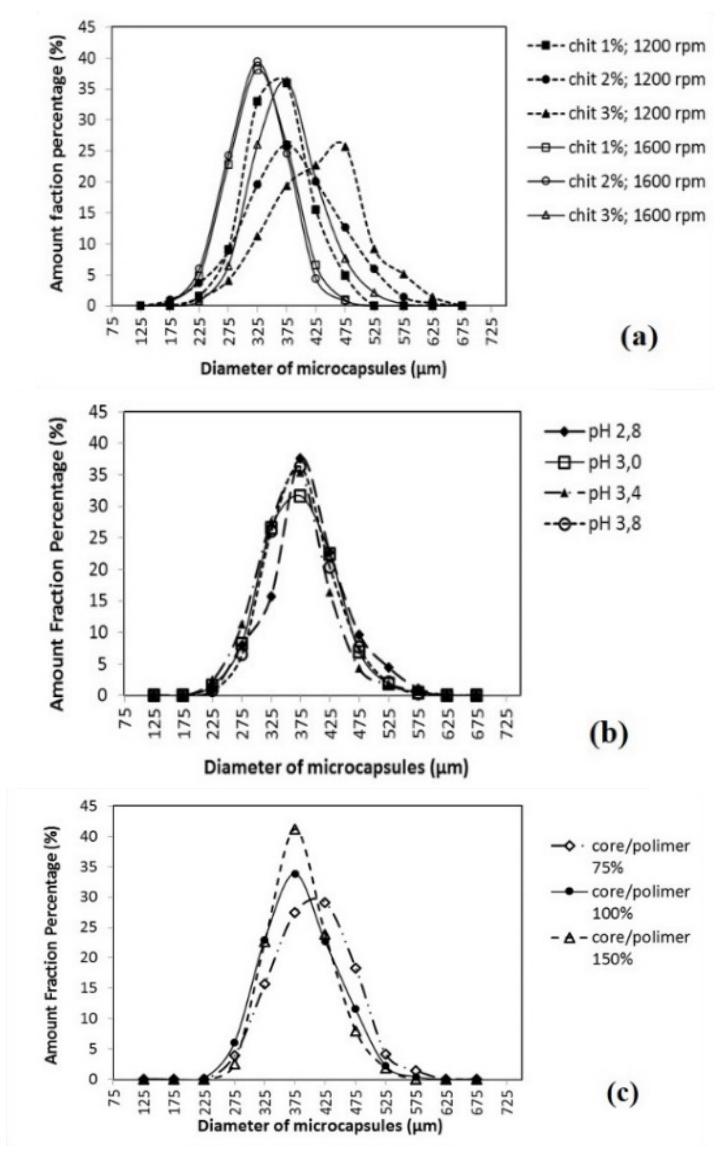
Figure 3 Particle size distribution of microcapsules with various: (a) polymer concentration and stirring intensity, (b) pH of polymer and cross-linker solution, (c) ratio of core to polymer concentration.
Generally, obtained encapsulation efficiencies were low which less than 25%. Some others research agreed with the result. Anthocyanin encapsulation efficiency obtained by using ionic gelation technique with chitosan-NaTPP was 33% [18]. While, other research obtained anthocyanin encapsulation efficiency of 23% with similar technique, carrier material and cross-linker [19]. The low encapsulation efficiency was caused by the weakness of chitosan-TPP complex, so that encapsulated anthocyanins were resisted from process after microencapsulation such as filtering, washing, and drying.
3.2 Influence of pH of chitosan and NaTPP solution
The influence of pH of chitosan and NaTPP solution varied at 2.8; 3.0; 3.4; 3.8; and 4.2 on mean encapsulation efficiency, mean diameter and antioxidant activity of microcapsules was showed at Table 2. Antioxidant activity was expressed as antiradical power (ARP). The influence of pH of chitosan and NaTPP solution on anthocyanin microcapsules was involved with anthocyanins structure mentioned in introduction, also ionization degree of chitosan and TPP charge number as can be seen at Figure 4 [20]. Overall, mean encapsulation efficiency difference was not significant. But there was difference among samples. It can be seen that pH 3.0 was significantly differ with pH 2.8, pH 3.8, and 4.2. In range pH 3.0-4.2, there was tendency that mean encapsulation efficiency decreased with increasing of pH.
Table 2 The influence of pH of chitosan and NaTPP solution on mean encapsulation efficiency, mean diameter and antioxidant activity.
| pH of chitosan and NaTPP solution | Mean encapsulation efficiency (%) | Mean Diameter (μm) | Antiradical Power (ARP) (mg of DPPH/mg of\nencapsulated anthocyanin) |
|---|---|---|---|
| 2.8 | 22.44b,c | 386.7 | 74.15 |
| 3.0 | 24.81a | 374.3 | 75.27 |
| 3.4 | 24.03a,b | 362.7 | 64.62 |
| 3.8 | \(22.79^{b,c}\) | 375.7 | 53.59 |
| 4.2 | 22.17° | 51.40 |
Note: Similar superscript indicate unsignificantly difference with p>0.05
According to Figure 4 in that pH range, ionization degree of chitosan were similar, but charge number of TPP was raised with increasing of pH, although the value was not significant. The increasing of charge number of TPP indicated the increasing of intermolecular crosslinking, further chitosan-TPP matrix density also increased. In the other hand, anthocyanins species presence was on flavylium cation and quinoidal base in the range pH 3.0-4.2 in which the amount of flavylium cation decreased with increasing of pH level. There was possibility that positive charge of flavylium cation would interact with TPP
anion forming complex. The increasing of pH level reduced the interaction, further it induced encapsulation efficiency reduction, although as has been mentioned that there was only little increasing of chitosan-TPP matrix density with increasing of pH.
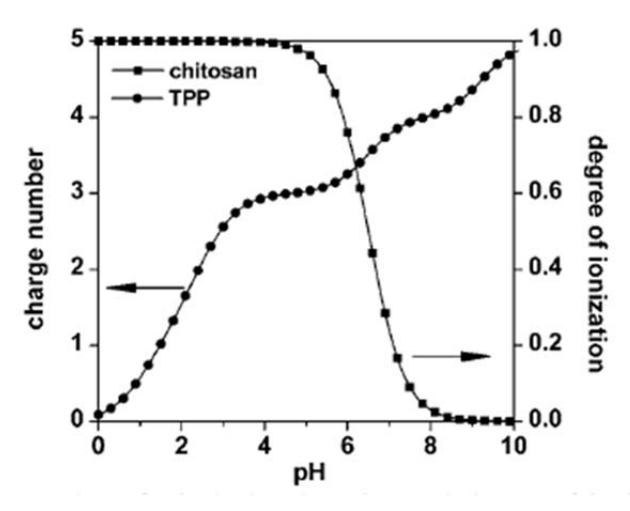
Figure 4 The influence of pH on ionization degree of chitosan and charge number of TPP.
However at pH 2.8, encapsulation efficiency was lower than that of pH 3.0 even the difference was significant. There was possibility that flavylium cation was superior than base quinoidal at pH 2.8. Flavylium cation competed with protonated amine group in interacting with negative charge of TPP. Furthermore, it reduced intermolecular chitosan-TPP crosslinking. Uncrosslinked chitosan would interact each other by self-assembly mechanism which obtained matrix density was weaker and encapsulation efficiency was reduced [21].
Antioxidant activity of microcapsules was in good agreement with obtained encapsulation efficiency. In range of pH 3.0- 4.2, antioxidant activity was lower with the increasing pH. That was caused by decreasing the amount of flaylium cation structure. The deviation was found at pH 2.8 in which antiradical power was lower compared with pH 3.0. It was known that higher anthocyanin concentration would increase antioxidant activity. Lower encapsulation efficiency indicated lower encapsulated anthocyanin concentration and further lowered antioxidant activity. But for similar encapsulation efficiency, antioxidant activity of pH 2.8 was higher than that of pH 3.8 and 4.2. It is because at pH 2.8 was dominated by flavylium cation, instead of pH 3.0 dominated by quinoidal base. Flavylium cation has higher anthocyanin stability than quinoidal base.
Mean diameter and particle size diameter were almost similar among microcapsules samples varied in pH 2.8; 3.0; 3.4; and 3.8. The results can be seen at Table 2 and Figure 3(b). In the range of pH variation, chitosan had been ionized completely so that viscosity of chitosan solution among samples were the same. In emulsification process, the obtained droplets sizes were similar if the fluids had same viscosity and they were given similar mechanical energy.
3.3 Influence of Ratio of Anthocyanins to Polymer Concentration
Ratio of anthocyanins to polymer concentration which was varied at 75; 100; and 150% (w/w) gave influence on microcapsule size, efficiency of encapsulation, and particle size distribution as can be seen in Table 3 and Figure 3(c). The smaller ratio of core to polymer concentration, the bigger mean microcapsule diameter. Particle size distribution of microcapsules was broader with decreasing ratio of core to polymer concentration. The result was involved with change of chitosan solution viscosity after anthocyanin extract was added. Used core in this research was differ with others research that was crude extract with Brix 83o , so there was water content in extract. Adding the core caused decreasing of system viscosity and furthermore the droplet obtained in emulsification was smaller if similar stirring intensity was given.
Table 3 The influence of ratio of anthocyanin to polymer concentration on mean encapsulation efficiency and mean diameter.
| Ratio of anthocyanin to polymer concentration | Mean encapsulation efficiency (%) | Mean diameter (µm) |
|---|---|---|
| 75% | 31.93 | 405.7 |
| 100% | 29.03 | 384.2 |
| 150% | 28.24 | 383.9 |
As can be seen in Table 3, obtained encapsulation efficiency was smaller with increasing ratio of core to polymer concentration. There was limitation of matrix polymer to entrap core material. Moreover, the more core indicated the more flavylium cation in internal phase. As had been mentioned before, flavylium cation competed with protonated amine group so it reduced chitosan-TPP cross-linking. Matrix density became weaker and anthocyanins encapsulation in chitosan matrix was declined.
3.4 Morphology of Anthocyanin Microcapsules
Morphology of microcapsules prepared with stirring intensity 1600 rpm, chitosan concentration 3% (w/v), ratio of core to chitosan concentration 150% (w/w) at pH 3.0 and observed by SEM can be shown at Figure 5. It can be seen that anthocyanin microcapsules had spherical form but was irregular. Microcapsules had pores and the surface was rough. That may be due to the formation of interconnection chain resulted from intermolecular chitosan-TPP bond. Similar trends were found on anthocyanin microcapsules prepared by chitosan-TPP ionic gelation technique [18], and heparin microcapsules produced by using emulsification-crosslinking technique [21].
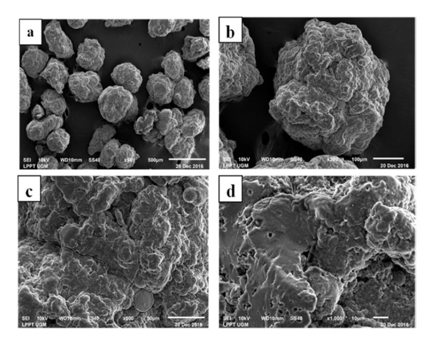
Figure 5 Morphology of anthocyanin microcapsule with magnification: (a) 50x, (b) 200x, (c) 500x and (d) 1000x.
3.5 Fourier Transform Infrared Spectrophotometry (FT-IR) Analysis
For predicting chemical bonding and interaction in anthocyanins microcapsules, FTIR spectroscopy was conducted for anthocyanins extract, chitosan, NaTPP, anthocyanin-loaded microcapsules, released anthocyanin-loaded microcapsules, as can be seen at Figure 6. Region with absorption bands around 3300-3450 cm<sup>-1</sup> indicated stretching vibration of hydroxyl group (O-H), amine (N-H), and carboxyl (C-H). Chitosan spectrum had band at 3357.2 cm<sup>-1</sup> but it was shifted to wavenumber 3448.2 cm<sup>-1</sup> at spectrum of anthocyanin-loaded microcapsules and also to wavenumber 3425.58 cm<sup>-1</sup> at spectrum of released anthocyanin-loaded microcapsules with both bands were broader than that of chitosan. The observation indicated that there were hydrogen bonding at anthocyanin-loaded microcapsule and also released anthocyanin-loaded microcapsule.
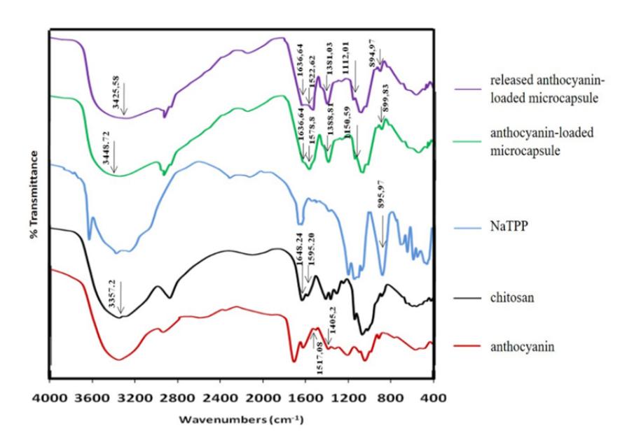
Figure 6 Fourier Transform Infrared Spectrophotometry (FT-IR) Spectrum.
Bands at 1500-1700 cm<sup>-1</sup> were related to stretching vibration of C=O, C=N, and C=C group. Peaks of chitosan spectrum at 1648.24 cm<sup>-1</sup> and 1595.20 cm<sup>-1</sup> were shifted to wavenumber 1636.64 cm<sup>-1</sup> and 1578.80 cm<sup>-1</sup> at spectrum of anthocyanin-loaded microcapsule and also shifted to wavenumber 1636.64 cm<sup>-1</sup> and 1522.62 cm<sup>-1</sup> at spectrum of released anthocyanin-loaded microcapsule. The shifting was related to chitosan and tripolyphosphate interaction. Chitosan spectrum had band at 1595.2 cm<sup>-1</sup> indicating N-H bending vibration and it was changed to 1578.80 cm<sup>-1</sup> at anthocyanin microcapsules and 1522.62 cm<sup>-1</sup> released anthocyanin-loaded microcapsules. They were related to protonated NH<sub>3</sub> [21].
Interaction of chitosan and TPP was also supported by shifting of chitosan band and peak intensity at 897.90 cm<sup>-1</sup> contributing N-H out of bending to wavenumber 899.83 cm<sup>-1</sup> at anthocyanin-loaded microcapsules and also to wavenumber 894.97 cm<sup>-1</sup> at released anthocyanin-loaded microcapsules. It was known that band of NaTPP spectrum at 895.97 cm<sup>-1</sup> referred to P-O-P asymmetric stretching, so possible interaction between chitosan and NaTPP was ionic interaction, exactly between protonated amine of chitosan and P-O group of TPP.
C-O-C alkyl group was assigned by peak of anthocyanin microcapsules at 1150.59 cm<sup>-1</sup>. It was shifted to wavenumber 1112.01 cm<sup>-1</sup> at released microcapsule spectrum. The difference may be due to interaction of anthocyanin and chitosan-TPP matrix. Specific C=C-C aromatic ring stretching
vibration of anthocyanin spectrum was shown by bands at 1517.08 and 1405. 2 cm<sup>-1</sup> and they were shifted to wavenumber 1522.62 and 1381.03 cm<sup>-1</sup> at released microcapsules spectrum, even though the shifting was overlap with stretching vibration of C=N or NH<sub>3</sub><sup>+</sup> group. Based on the FT-IR analysis, the hypothesis of interaction among anthocyanin, protonated chitosan, and tripolyphosphate ion can be depicted by Figure 7.
Figure 7 The hypothesis of interaction among anthocyanin, protonated chitosan, and tripolyphosphate ion.
4 Conclusions
Anthocyanins microcapsules were successfully prepared by emulsification-crosslinking technique in which water in oil (W/O) emulsion was formed. In the process, chitosan having role as polymer (carrier material) interacted ionically with NaTPP as cross-linker. The parameters influencing microencapsulation were studied and optimized. Parameters producing better microcapsules were stirring intensity 1600 rpm, chitosan concentration 3% (w/v), pH of chitosan and NaTPP solution 3.0, ratio of core to chitosan concentration 150% (w/w). Morphology of microcapsules was sphere but irregular. By FT-IR analysis, it was obtained that there was bonding and interaction among anthocyanin, chitosan, and NaTPP.
Acknowledgement
This research was supported by Fellowship of Ministry of Research, Technology and Higher Education of the Republic of Indonesia 2014/2016.