
1 Introduction
Dental caries is a risk factor for systemic disease. Dental caries bacteria have been found in coronary atherosclerotic plaque specimens of patients who died of a heart attack [1,2]. This shows the importance of preventing dental caries.
The National Health Research (Riskesdas) 2007 reported caries active prevalence in Indonesia at 43.4%. 14 provinces have caries prevalence above national prevalence.Basic health research conducted in 2007 and 2013 showed that the rates for need of dental health were 23.5% and 25.9% [3,4]. Of the substances for treatment of dental caries on the market today, many have side effects such as allergies and they are expensive because as yet they must be imported from abroad. Therefore, an immunomodulatory substance is needed that can be accepted by the body with minimal side effects. One solution is to create materials derived from natural materials, for example from coffee beans.
Coffee may protect against periodontal diseases [5]. Coffee contains bioactive components such as flavonoids, xanthine, antioxidants and alkaloids. These are polyphenols with anti-inflammatory and antibacterial activity and can prevent platelet aggregation [6-8]. This has been attributed to the mechanism of one of the proteins, which can help in the prevention, inhibition and treatment of such diseases. The protein content of robusta and arabica coffee beans is in the range of 10-13%. Coffee has an inhibitory effect on S. mutans, inhibits S. mutans adhesion and whitens teeth, while it is known that black coffee prevents cavities [9-12].
Caries can be prevented and restored by modulating the immune response of the sufferer. The dental caries immune response produces among others, IL-1β, IL-1α, and TNF-α. Previously our team has proven that robusta coffee can increase the expression of IL-1β and TNF-α, which stimulate phagocytosis in vitro of S. mutans and decrease inflammatory cells (monocytes, lymphocytes, neutrophils) in vivo in treatment of dental caries [13-15]. Robusta is also thought to affect fibroblast cells and IL-1α expression. IL-1α is a dual-function cytokine, meaning that in addition to functioning as a classical cytokine via cell surface receptor ligation, full-length IL-1α can also directly regulate gene expression. As a prototypical proinflammatory cytokine, IL-1 drives local and systemic inflammation after injury and is critically involved in the pathobiology of immune and inflammatory conditions. IL-1, IL-6, and b-FGF (basic fibroblast growth factor) may be produced in vivo by residual lens epithelial cells (LECs), causing post-operative inflammation and cultured LEC proliferation after cataract surgery [16-18]. Fibroblasts are cells that are oval, large and pale, with fine chromatin and a clear core nucleolus with many irregular cytoplasmic branches. Fibroblasts play an important role in the healing process. Various growth factors strongly support fibroblast proliferation, for example for healing wounds involving reepithelialization, granulation tissue formation, reduction of the inflammatory process, interleukin IL-1 expression [19,20].
2 Materials and Methods
2.1 Animal Model
64 male Wistar rats weighing 100-200 gr BW, at 2-3 months old, were obtained from the Animal House at Biomedic laboratory Faculty of Dentistry Universitas Jember. The rats were kept in separate cages in a well-ventilated room at standard experimental conditions. Approval from the Gajah Mada University animal ethics committee was obtained for this project.
2.2 Robusta Coffee
Robusta coffee bean paste was made by mixing the basic paste extract (magnesium carbonat, calcium carbonat, glycerine, TEA (triethanolamine), propylene glycol, and aquadest).
2.3 Research Groups
The animals were divided into 4 groups and 3 subgroups (7-, 14-, 21-day rats) with each group containing 4 animals; in the treatment groups, perforations were made in the teeth to direct pulp capping using robusta coffee. Control Group: untreated rats; 25% Coffee Group: cavities + treated with 25% coffee bean paste; 50% Coffee Group: cavities + treated with 50% coffee bean paste; 75% Coffee Group: cavities + treated with 75% coffee bean paste. The 7-, 14-, 21-day rats were sacrificed serially after which their teeth were prepared for analysis of the number of dental pulp fibroblasts by HE staining and IL-1α expression by immunohistochemistry.
2.4 Immunohistochemistry Methods
Mixing was done 3 times for deparaffinization using xylol. The xylol was eliminated with absolute ethanol ranging up to 70%, the last added with water, after which the mixture was washed with PBS pH 7.4. Debris was removed with 0.025% trypsin. The mixture was flooded with 3% H2O2 solution for 10 minutes and then washed 2 times with PBS, after which blocking was carried out with 3% BSA for 10 minutes. Reacted with rat IL-1α antibody (Dako) and incubated for 24 hours at a temperature of 40 °C in a humidity chamber. Reacted with biotylized secondary Ab (goat anti-rat IL-1α, Dako) for 1 hour and then washed 3 times with PBS every 5 minutes. Added with peroxidase and labeled streptavidin, incubated for 1 hour, washed 3 times with PBS, and treated with DAB (Dako), creating a new substrate. Incubated for approximately 30 minutes at room temperature with shaking. Washed with distilled water and added with Meyer-HE for 10 minutes. Washed with tap water and then with distilled water. The preparations were dried, added with entelan, and covered with a coverglass. Microscopic analysis was done under a light microscope with 400x magnification, which was analyzed over 3 fields of view. The parameter was the amount of leucocytes, which express IL-1α. The data were analyzed descriptively and by ANOVA, followed by an LSD test.
2.5 HE Staining Method
The dental tissue was inserted in formalin, decalcified, deparanized, paraffin blocked, immersed and washed with PBS. It was cut with microtome at 4-6 micron and attached to a glass slide. Then staining with haematoxilin eosin (HE) was done. Assessment and analysis of the fibroblasts was done under a light microscope with 400x magnification over 3 fields of view. The data were analyzed by ANOVA, followed by an LSD test.
3 Results and Discussion
3.1 Effect of Robusta Coffee on IL-α Expression
Figures 1 and 2 show that robusta coffee decreased IL-1α expression in the dental pulp. Cells expressing IL-1α are brown (black arrow). Descriptive analysis under a light microscope with 400x magnification revealed that the more days (7, 14, 21 days), the more IL-1α expression decreased in the dental pulp. The control group's IL-1α expression decreased the most, because it was untreated, while IL-1α was produced physiologically. This was confirmed by the ANOVA results followed by the LSD test, in which there were significant differences (p ≤ 0.05).
High protein content or bioactive content in robusta coffee are suspected to be immunogenic or act as immunomodulator, thus allegedly having the ability to improve the immune system. This study proved that robusta coffee decreased the expression of IL-1α either inside the cell or outside the cell. It is suspected that immunomodulatory proteins potentially work against lymphokines produced by immunocompetent cells such as IL-1α. The decrease of IL-1α is thought to be due to the content of flavonoids, xanthine, chlorogenic acid, and alkaloids in robusta coffee. Flavonoids act as anti-inflammatory agent, analgesic, and antioxidant [21]. Some flavonoid compounds may inhibit the release of arachidonic acid and lysosomal enzyme secretion from the membrane by blocking the cyclooxygenase and lipoxygenase pathways, thus decreasing the levels of prostaglandins and leukotriene (inflammatory mediators), for which one of the inflammatory mediators is IL-1α [22].
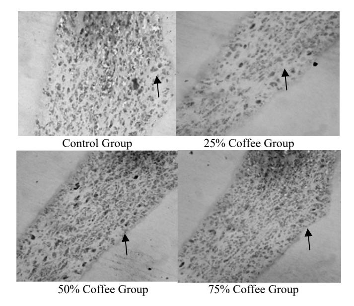
Figure 1 IL-1α expression in Wistar rat dental pulp (black arrow).
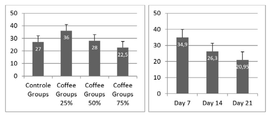
Figure 2 Bar chart of IL-1α expression in Wistar rat dental pulp
The results of this study indicate that the control group had the least fibroblasts. The robusta coffee group with the highest number of fibroblasts was the 75% Coffee Group (Figures 3 and 4). Robusta coffee as pulp capping material reduces inflammation and speeds up the healing process. This is caused by IL-1α that is synthesized and released by macrophages and plays an essential role in all inflammatory processes. Therefore, if IL-1α decreases, the inflammatory process is reduced and the healing process increases, for example proliferation of fibroblasts [9]. IL-1α also is made by B cells, T cells, large granular lymphocytes, endothelial cells, astrocytes, muscle cells, keratinocytes and many types of fibroblasts. IL-1α may inhibit the reparative function of fibroblasts by stimulating their proliferation and synthesis of collagen and tissue inhibitor of metalloproteinase (TEAM) [22].
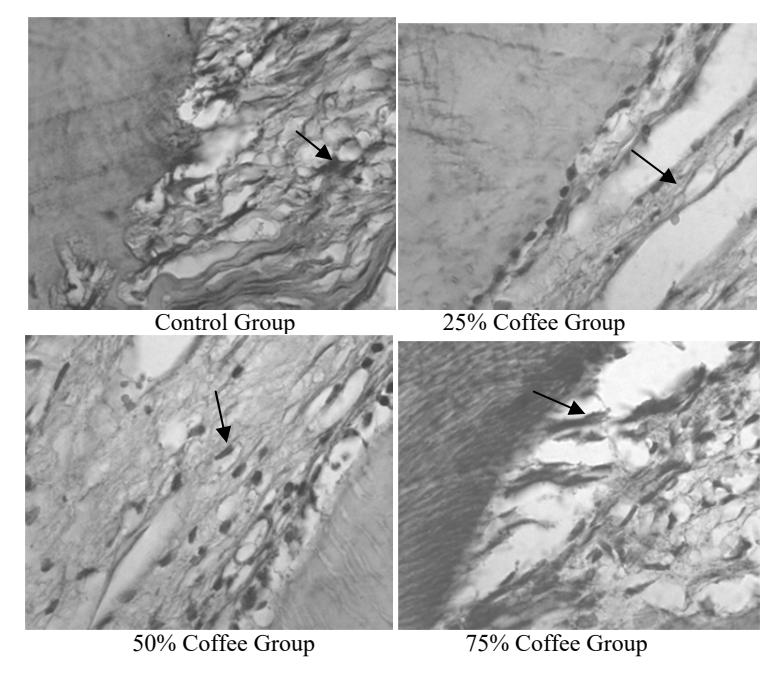
Figure 3 Fibroblasts in Wistar rat dental pulp (black arrow).
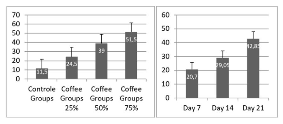
Figure 4 Bar chart of the number of fibroblasts in Wistar rat dental pulp.
3.2 Supporting Data (number of leukocytes)
Other data that could support this study are the number of inflammatory cells, which decreased from day 7 and 14 to 21 (see our previous work), where on the 7th day neutrophils and many macrophage cells and lymphocytes were visible. On day 14 there was more domination of macrophage cells and lymphocytes, while the number of neutrophils was very small. On day 21 there were still macrophages and lymphocytes, but the number of fibroblasts was higher (5) [15]
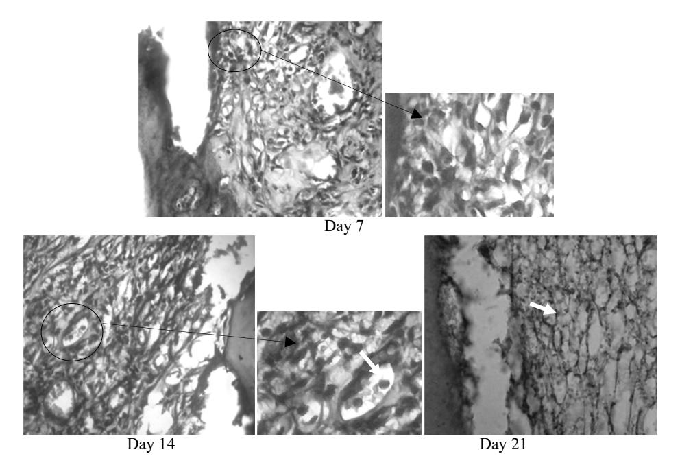
Figure 5 Inflammatory cells in Wistar rat dental pulp in the coffee groups.
4 Conclusion
Robusta coffee bean paste as pulp capping material increases the healing process in Wistar rat dental pulp.
Acknowledgements
A big thank you to Research University of Jember, which gave an opportunity to obtain research grants and RISTEK DIKTI, which has provided funding for this study.