
1 Introduction
Plants are utilized all around the globe. Native communities utilize flowering plants to cure many ailments. However, only few are known to modern science [1]. In recent years, the search for novel antimicrobial agents in plants has increased [2]. These antimicrobial agents, such as tannins, alkaloids and flavonoids, are abundantly present in plants [3]. They are cost-effective, easily available and relatively safe [4]. There are numerous findings on the antimicrobial potential of various herbal dilutions [5-7].
Monotheca buxifolia belongs to the Sapotaceae family of flowering plants. This plant species is native to Pakistan, Afghanistan, Oman and Saudi Arabia. It is one of the imperative members of Pakistani forests, especially in the northern hilly areas. It is mostly used for fuel, building shelters and fences around farms and fields. Its fruit is sold in markets providing a source of money for the local people [8]. In traditional recipes, the fruit of M. buxifolia is consumed as a laxative, cleansing agent, vermicide, antipyretic, and for the treatment of gastrointestinal ailments [9-12]. The leaves are a rich source of anthraquinones, flavonoids, terpenoids, glycosides, saponins, glucose, tannins and polyphenolic residues. These compounds have strong anti-inflammatory and pain-relieving properties [11]. The fruit of the plant also has a high antioxidant level [13]. Furthermore, M. buxifolia fruit extract also exhibits inhibitory activity against urease enzymes [14].
The Sapotaceae family has been extensively screened for antimicrobial [15,16], antipyretic [17], CNS sedative[18], anti-inflammatory [19,20], anthelmintic [21] and anti-nociceptive [22] potential in numerous in vitro and in vivo research trails. In this study, we tested petroleum ether, n-hexane and methanolic extracts of the aerial parts of M. buxifolia for antibacterial, antifungal, cytotoxic and antioxidant potential.
2 Materials and Methods
2.1 Plant Collection
M. buxifolia is a small thorny tree or shrub that belongs to the Monotheca genus. It is an important plant species and exists in dominance in a number of forest areas in the Dir district, Pakistan (Figure 1).
Figure 1 Monotheca buxifolia stem, fruit, leaves and spines.
A sample was also deposited at the herbarium of the University of Peshawar. The plant materials were washed, cleaned and shade dried. After drying, the samples were ground to powder by electric blender.
2.2 Extraction of Plants Material
After air-drying, 100 g of each sample (fruit, leaves and seeds) was added to 300 mL of n-hexane, petroleum ether and methanol for fifteen days at room temperature. These extracts were filtered via filter paper (Whatsman) and the filtrate was concentrated at 40 °C under vacuum using a rotary evaporator to recover the solvent. The filtrates were stored in a freezer at 4 °C until use in later biological assays.
2.3 Anti-Bacterial Screening
Bacterial strains were provided by the Department of Biotechnology, University Peshawar, Pakistan. The strains were identified by various biochemical tests [23]. The different fractions of the plant extracts were screened for possible antibacterial activity by agar well diffusion [24]. During preparation for inoculation, the tested bacteria were cultured in nutrient broth at 37 °C for 24 hours. 30 μl of prepared culture was spread on the surface of nutrient agar media to test for bacterial pathogens. After inoculation, the culture was transferred to plates. Wells (6 mm) were made with a sterile borer, keeping a distance of 24 mm between two adjacent wells. About l00 µl of 3 mg/mL of the respective extracts dissolved in DMSO was added to the wells. Other wells were supplemented with DMSO and 10 µg Amoxicillin, Ciprofloxacin, and Rifampicin to serve as positive and negative controls.
The plates were then incubated for 24 hours at 37 °C, after which they were observed for zones of inhibition. All experiments were conducted in triplicate. The percent zone of inhibition was measured in comparison with the positive control using the following equation:
% Inhibition = \[\left(\frac{\text{Zone of Inhibition of Sample}}{\text{Zone of Inhibition of Standard}}\right) x 100\] (1)
2.4 Antifungal Screening
Fungal strains, i.e. Aspergillus niger, Aspergillus parasiticus, Penicillium notatum, Fusarium oxysporum and Verticilium, were used to determine the antifungal potential of the plant extracts. The fungal strains were provided by COBAM, University of Peshawar. For the growth of the fungi, 4 mL Sabouraud dextrose agar media was prepared in distilled water. After cooling, 67 µl of test sample was added to the test tubes containing non-solidify SDA media.
The test tubes were kept in a slanted position, cooled and incubated for 24 hours at 28 ± 1 °C to check sterility. After 24 hours, each labeled test tube was inoculated with a piece of inoculum removed from a seven-day old fungal culture and incubated at 25 ± 1 °C for 7 days in an incubator. Miconazole antibiotic and DMSO were used as positive and negative control respectively. During incubation, the cultures were observed twice weekly. The results were noted on day 7 by measuring the linear growth (mm) in the slanted test tubes loaded with sample in comparison with Miconazole (negative control), after which the percent inhibition of fungal growth was calculated by using the following equation:
% Inhibition = \[\left(\frac{\text{Linear growth in test (mm)}}{\text{Linear growth in standard (mm)}}\right) x 100\] (2)
2.5 Antioxidant Activity/Nitric Oxide (no) Free Radical Scavenging Assay
The nitric oxide radical scavenging activities were described according to Makhija [25] with some modification. In aqueous solution at normal pH (7.2), sodium nitroprusside decays into nitric oxide radicals. In the presence of oxygen, these radicles react with oxygen, leading to the production of steady intermediates (nitrate and nitrite), which can be noted via Griess reagent [26]. 70 µl of sodium nitroprusside solution was added to 10 µl of each dilution at variable concentrations (100-600 µg/mL). The blend was then incubated at room temperature for 110 min. After incubation, 50 µl of the reacting mixture was blended with Griess dilution (1% sulphanilamide, 1% Nnapthylethylenediamine hydrochloride in 2% o-phosphoric acid). The absorbance was noted at 546 nm against blank. The scavenging percentage was calculated using the following equation:
% scavenging = \[\left(\frac{\text{absorbance of control} - \text{absorbance of sample}}{\text{(absorbance of control}}\right) x 100\] (3)
2.6 Cytotoxicity/Brine Shrimp Lethality Bioassay
The cytotoxic potential of different extracts was determined using brine shrimp lethality bioassay by following the method of Ullah [27]. Different concentrations (5, 50, 500 μg/mL) of plant extract were prepared in methanol, ether and n-hexane. After 48 hours of hatching Artemia salina and maturation as nauplii the larvae were transferred using a Pasteur pipette. Ten larvae were transferred to each vial. The vials under illumination were then placed at a temperature of 25-27 °C for 24 hours. Vials were filled with brine solution for negative control and cytotoxicity, while Etoposide (7.4625 µg/ml) was used for positive control. After 24 hours of incubation, the numbers of live and dead brine shrimps were counted using a magnifying glass. Finally, a Probit analysis was used to determine and analyze the LD50 values with 95% confidence intervals.
2.7 Statistical Analysis
Statistical analysis was performed using Microsoft Excel (2013) and Graph Pad (v 5.0). All experimental results were expressed as means ± SD of triplicate.
3 Results
3.1 Antibacterial Activity
The majority of the tested bacterial strains were vulnerable to antibiotics, as shown in Figure 2. No bacterial strain was found to be resistant to any antibiotics tested.
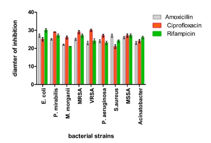
Figure 2 Exposure design for exposing bacterial strains to standard antibiotic discs.
3.2 Antibacterial Disk Diffusion Assay
The pet-ether fruit extract was most active against P. mirabilis, MSSA and M. morganii with inhibitory zones of 23 ± 0.89, 21.2 ± 1.02 and 20 ± 0.76 mm respectively. The methanolic fruit extract was most active against VRSA, S. aureus and Acinetobacter (Table 1). The n-hexane leave extract was most active against Acinetobacter and M. morganii. The seed extract showed no activity (Table 2). The methanolic seed extract had the highest activity against VRSA, MRSA and Acinetobacter with inhibitory activity at 17 mm, 15 mm and 14 mm respectively (Table 3).
Table 1 Antibacterial activity of leaves, fruit and seeds using ether as solvent.
| Extract | Acinetoba | E. coli | P.mirab | MSSA | MRSA | P.aerugin | S. aureus | M.morg | VRSA |
|---|---|---|---|---|---|---|---|---|---|
| cter | ilis | osa | ani | ||||||
| Leaves | 13±0.29 | 13±0.00 | 12±0.19 | 1±0.14 | 13±0.5 | 1±0.00 | 15±0.71 | 11±0.00 | 15±0.12 |
| Fruit | \(16\pm0.57\) | \(14\pm0.00\) | 23±0.55 | \(21\pm0.0\) | \(18\pm0.08\) | \(11\pm0.57\) | 14.5±1.3 | \(18\pm0.98\) | 11±0.32 |
| Seeds | 0 | 0 | 0 | 0 | 0 | 0 | 0 | 0 | 0 |
| Amoxil | 23±0.87 | \(27\pm0.98\) | 25±0.45 | 26±640 | \(25\pm0.68\) | \(22\pm0.86\) | \(27\pm0.87\) | 22±0.35 | \(23\pm0.98\) |
Table 2 Antibacterial activity of leaves, fruit and seeds using n-hexane as solvent.
| Extract | Acinetoba | E. coli | P.mirab | MSSA | MRSA | P.aerugin | S. | M.morgan | VRSA |
|---|---|---|---|---|---|---|---|---|---|
| cter | 2.00. | ilis | 1120012 | osa | aureus | i | |||
| Leaves | 13±0.00 | 14±0.10 | 13±0.79 | 12±0.19 | 10±0.32 | 12±0.31 | 12±0.31 | 13±0.37 | 13±0.00 |
| Fruit | \(17\pm0.00\) | \(12\pm0.80\) | \(13\pm0.71\) | \(13\pm0.00\) | \(13\pm0.01\) | \(12\pm0.14\) | \(13\pm0.78\) | \(13\pm0.12\) | \(14\pm0.57\) |
| Seeds | \(12\pm0.56\) | \(11\pm1.10\) | \(10\pm0.13\) | 0 | 0 | \(14\pm0.91\) | \(11\pm0.00\) | \(2\pm0.10\) | \(1\pm0.00\) |
| Amoxil | \(23\pm0.87\) | \(27\pm0.98\) | 25±0.45 | 26±640 | 25±0.68 | \(22\pm0.86\) | \(27\pm0.87\) | 22±0.35 | \(23\pm0.98\) |
Table 3 Antibacterial activity of leaves, fruit and seeds using methanol as solvent.
| Extract | Acinetoba cter | E. coli | P. mirabilis | MSSA | MRSA | P. aeruginosa | S. aureus | M.morgani | VRSA |
|---|---|---|---|---|---|---|---|---|---|
| 14.0.00 | 14:12 | 12:0.71 | 14:014 | 10.071 | |||||
| Leaves | \(13\pm0.00\) | \(14\pm0.82\) | \(1\pm0.0\) | 14±1.2 | \(12\pm0.71\) | \(13\pm0.12\) | \(13\pm0.81\) | \(14\pm0.14\) | \(13\pm0.71\) |
| Fruit | 16±1.20 | \(14\pm0.00\) | \(14\pm0.12\) | \(11\pm0.00\) | \(12\pm0.54\) | \(11\pm0.57\) | \(15\pm0.32\) | \(11\pm0.90\) | \(22\pm0.00\) |
| Seeds | \(14\pm0.55\) | 0 | 0 | \(13\pm0.16\) | \(15\pm0.45\) | \(13\pm0.17\) | \(14\pm0.87\) | 0 | 17±1.3 |
| Amoxil | 23±0.87 | \(27\pm0.98\) | 25±0.45 | 26±640 | \(25\pm0.68\) | 22±0.86 | \(27\pm0.87\) | 22±0.35 | 23±0.98 |
3.3 Antifungal Disc Diffusion Assay
The methanolic fruit extract showed no activity against any fungi, while the pet-ether fruit extract showed low activity against P. notatum and F. oxysporum (Figure 3 and Table 4).
Figure 3 Fixed oil extracted with n-hexane showed inhibition against (a) A. parasiticus, (b) F. oxysporium, (c) H. mycelia, and (d) Verticilium.
| Table 4 Antifungal activity of leaves, fruit and seeds using ether as solvent. | |||||
|---|---|---|---|---|---|
| Part of plant | A. parasiticus | A. niger | P. notatum | F.oxysporum | Verticilium |
Leaves - - - 40±3.98 - Fruit - - 37±2.56 17±2.14 - Seeds - - - 54±4.68 - Positive control 55±4.09 50±5.00 59±3.23 65±7.02 56±0.00
Values are expressed as means ± standard deviation (n = 3) – no activity.
The methanolic seed extract showed 55% inhibition against F. oxysporum while showing no activity against the rest of the fungi. The n-hexane fruit and seed extract showed antifungual activity against P. notatum and F. oxysporum respectively (Table 5).
Table 5 Antifungal activity of leaves, fruit and seeds using n-hexane as solvent.
| Part of plant | A. parasiticus | A. niger | P. notatum | F.oxysporum | Verticilium |
|---|---|---|---|---|---|
| Leaves | - | - | - | - | - |
| Fruit | - | - | 8±0.00 | - | - |
| Seeds | - | - | - | 59±6.00 | - |
| Positive control | 55±4.09 | 50±5.00 | 59±3.23 | 65±7.02 | 56±0.00 |
Values are expressed as means ± standard deviation (n = 3) – no activity.
The methanolic fruit extract showed 8% inhibition against P. notatum and no activity against A. niger, A. parasiticus, F. oxysporium and Verticilium (Table 6). Similarly, the pet-ether leaf extract showed 40% inhibition against F. oxysporum. The crude methanolic leaf extract showed no activity against A. niger, A. parasiticus, F. oxysporium, P. notatum and Verticilium (Table 4).
Table 6 Antifungal activity of leaves, fruit and seeds using methanol as solvent.
| Part of plant | A. parasiticus | A. niger | P. notatum | F.oxysporum | Verticilium |
|---|---|---|---|---|---|
| Leaves | - | - | - | - | - |
| Fruit | - | - | - | - | - |
| Seeds | - | - | - | 55±5.36 | - |
| Positive control | 55±4.09 | 50±5.00 | 59±3.23 | 65±7.02 | 56±0.00 |
Values are expressed as means ± standard deviation (n = 3) – no activity.
3.4 Cytotoxic Activity
The methanolic extracts of fixed oil, seeds and leaves showed 80% inhibition at 5 µg with LD50 at 0.429 (Table 7). The pet-ether fruit extract had the highest cytotoxic activity, causing 60% of lethality in Artemia salina, with LD50 at 3.307 (Table 8). The methanolic extracts of fruit, seed and fixed oil also showed 90% of lethality in Artemia salina.
Sample Dose (µg/mL) Total larvae Survivors Inhibition % LD50 Fruit 5 µg 10 1 90 50 µg 10 0 100 1.710 500 µg 10 0 100 5 µg 10 2 80
50 µg 10 1 90 0.429
500 µg 10 0 100
Table 7 Average mortality of Artemia salina larvae out of 10 at several concentrations using methanol as solvent.
| Table 8 | Average mortality of Artemia salina larvae out of 10 at several | |||||
|---|---|---|---|---|---|---|
| concentrations using ether and n-hexane as solvent. |
Seeds (methanol) 5 µg 10 2 80 0.429
| Sample | Dose (µg/mL) | Total larvae | Survivors | Inhibition % | LD50 |
|---|---|---|---|---|---|
| 5 µg | 10 | 4 | 60 | ||
| Fruit (ether) | 50 µg | 10 | 2 | 80 | 3.307 |
| 500 µg | 10 | 0 | 100 | ||
| 5 µg | 10 | 2 | 80 | ||
| Leaves | 50 µg | 10 | 1 | 90 | 0.429 |
| (n-hexane) | 500 µg | 10 | 0 | 100 |
3.5 Antioxidant Activity
Fixed oil
The scavenging activity was highest in the methanolic leaf extract at 32.1, 40.38, 49.1, 57.5, 66.4 and 75.3% for concentrations of 100, 200, 300, 400, 500 and 600 µl respectively (Figure 4).
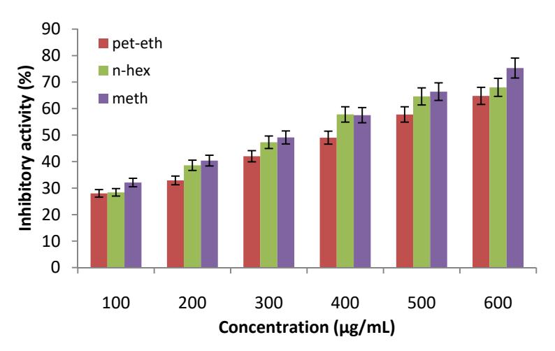
Figure 4 Nitric oxide (NO) free radical scavenging assay (anti-oxidant activity) of M. buxifolia leaf extract.
For the n-hexane fruit extract, the scavenging activity was 25.18%, 34.85%, 42.88%, 50.05%, 55.65% and 64.96% at concentrations of 100, 200, 300, 400, 500 and 600 µl respectively (Figure 5). For the pet-ether fruit extract it was 30.36%, 39.7%, 49.2%, 58.1%, 66.3% and 73.2% at concentrations of 100, 200, 300, 400, 500 and 600 µl respectively. For the fixed oil extract it was 23.32%, 31.69%, 35.21%, 42.12%, 54.23% and 63.45% at concentrations of 100, 200, 300, 400, 500 and 600 µl respectively (Figure 6). The methanolic seed extract showed the highest scavenging activity against nitric oxide (NO) radicals (Figure 7).
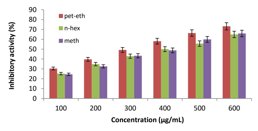
Figure 5 Nitric oxide (NO) free radical scavenging assay (anti-oxidant activity) of M. buxifolia fruit extract.
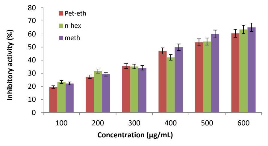
Figure 6 Nitric Oxide (NO) free radical scavenging assay (anti-oxidant activity) of M. buxifolia fixed oil extract.
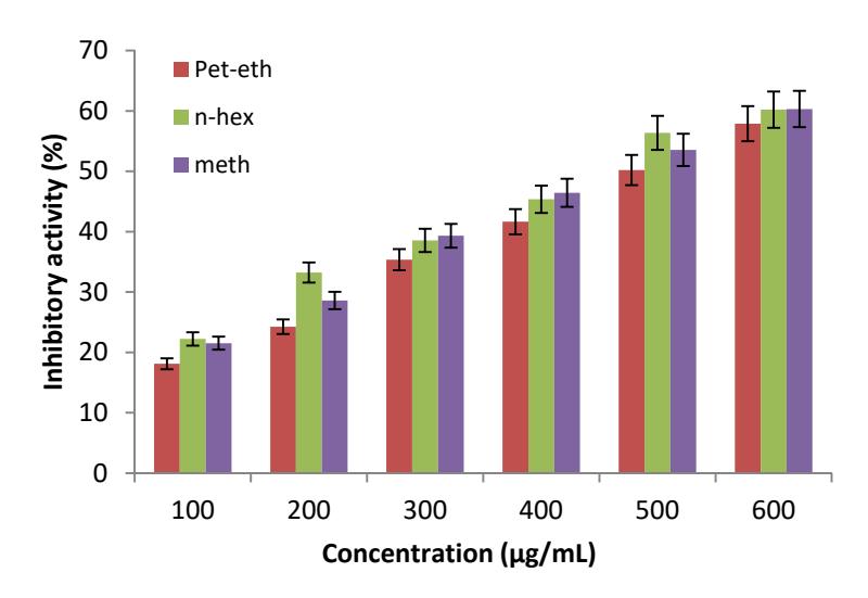
Figure 7 Nitric oxide (NO) free radical scavenging assay (anti-oxidant activity) of M. buxifolia seed extract.
4 Discussion
Antibiotic resistance levels are rising, therefore the search for novel bactericidal agents has increased over the past years. A main focus is using medicinal herbs that are widely available, have fewer side effects and have excellent antimicrobial potential [28]. Hence, the investigation of substitute medicines from plants that are active against multi-resistant pathogenic bacteria has increased all around the globe [29]. Currently, against multi-drug resistant bacteria such as VRE, MRSA, VRSA and P. aeruginosa mostly plants are used, having excellent antimicrobial potential [30-35].
Each plant extract that was tested in the present study exhibited antibacterial activity against the nine tested bacterial strains. However, differences were observed between the antibacterial potential of each extract. These differences can be attributed to differences in the chemical composition of these extracts as the secondary metabolites of plants have many effects, including antibacterial and antiviral activities [36,37]. Our results showed that Gram-positive bacteria were more sensitive to the studied plant extracts than Gram-negative ones. Our results are also supported by the previous study of Nair & Chanda [38]. Among the tested Gram-negative bacteria, Acinatobacter was found to be the most sensitive, while Morganella morganii was the most resistant bacteria. In the case of Gram-positive bacteria, MSSA was the most sensitive, while MRSA was the most resistant strain. Our results support the results reported by Mahesh & Satish [39]. The antifungal activity was not very notable compared to the antibacterial activity. Antifungal activity usually contrasts with antibacterial activity [40].
Antioxidants are effective against many disorders [41]. Our results showed that the methanolic leaf extract had the highest antioxidant potential. Numerous phytochemical constituents, such as flavonoids, phenyl propanoid and phenolic acids, have been recognized to be accountable for the antioxidant potential of plants [42]. The most important anti-oxidative components in M. Buxifolia are phenolic and flavonoids [13]. Our results are in contrast with Rehman, Khan [11], which may be due to differences in the extracts or process used. Cytotoxic activity was highest in the pet-ether fruit extract. Meanwhile, the methanolic extracts of fixed oil, seeds and leaves showed moderate cytotoxic activity, in accordance with Ullah and Ibrar [27].
It can be concluded that the plant M. buxifolia possesses significant antibacterial, antioxidant and cytotoxic activity and low anti-fungal activity. Further studies, regarding isolation and purification of new biomolecules, can show the detailed potential of the plant for use in anti-aging and anti-cancer medicine and novel antibiotics.
Acknowledgments
The authors thankfully acknowledge Mr. Ghulam Jelani, botanist for identification of the plant. The authors also wish to acknowledge Mr. Osama Ahmad & Arsalan Ahmad for kindly providing the dried plant material.