
1 Introduction
Spirulina platensis, which contains protein and vitamin, is widely used as a drug and food supplement in many countries [1]. It is also used as a natural dye for food and cosmetics [2]. The benefits come from pigment components contained within Spirulina, which are carotenoids, chlorophyll, and phycobiliprotein. Phycobiliproteins are accessory photosynthetic pigments that participate in an extremely efficient energy transfer chain in photosynthesis. The three main groups of phycobiliproteins are phycocyanins, allophycocyanins and phycoerythrins [3]. C-Phycocyanin (C-PC), a blue pigment (cyan means blue), absorbs light at a wavelength of 580-620 nm. It can be extracted from cyanobacteria such as Spirulina platensis and is widely used as a natural blue pigment in the food, cosmetical and pharmaceutical industries [4]. Recent studies have demonstrated that C-PC plays a role in antioxidants [5,6] and is a free radical scavenger, while it is also used as a fluorescent marker in biomedical research [7]. Liu, et al. [8] have reported that C-phycocyanin of Spirulina platensis inhibits K562 human leukemia cells. In Japan and China
phycocyanin is used as a natural coloring agent for food products, candy, jelly, ice cream and it is alo used in soft drinks such as Pepsi blue and in cosmetics for lipstick, eyeliner and eyeshadow [9]. Because of these benefits, several researchers have focused on developing efficient processes for mass production of phycocyanin-producing strains and extraction of phycocyanin from microalgae [10]. A number of factors can influence phycocyanin extraction. The most important ones are the cellular disruption method, the type of solvent, the biomass-solvent ratio, the extraction temperature and extraction time [11,12].
In this work, two different physical cell disruption methods for phycocyanin extraction from S. platensis were investigated. Moreover, the effects of solvent type on the phycocyanin yield and purity of the extracts were evaluated to establish the optimum conditions for phycocyanin extraction, followed by purification using ammonium sulfate precipitated and chromatography gel methods and characterization using SDS PAGE and spectrophotometry infrared.
2 Material and Methods
2.1 Optimization of Extraction Method
The dried cells of Spirulina platensis used in this study were obtained from BBPBAP (Balai Besar Perikanan Budidaya Air Payau) Jepara. C-PC was extracted from dried cells using physical methods, i.e. freeze-thawing and sonication for cellular disruption with different types of solvents, i.e. 0.01 M phosphate buffer pH 7, 0.01 M acetate buffer pH 5, water, and 1% CaCl2 solution. The freezing-thawing method was performed by homogenization with 1 portion of dried cells and 25 portions of solvent in an orbital shaker at room temperature for 24 h. After homogenization, the sample was put into a chiller for 1 h and then into a freezer for 20 h. The frozen solvent cells were left at room temperature for thawing.
Subsequently, an aliquot was centrifuged and the C-PC extract was collected for purity, phycocyanin concentration and yield analysis. The absorbance of the UV-Vis spectrum of the extracts was recorded with a UV-Vis spectrophotometer at wavelengths of 280 nm, 615 nm, 620 nm and 652 nm. The purity was evaluated according to absorbance ratio A620/A280. The concentration of phycocyanin (mg. mL-1) and the yield were determined by the formulas in Eqs (1) and (2), as described in Bennet and Bogorad [3].
\[C-PC (mg mL^{-1}) = \frac{A_{615} - (0.474 x A_{652})}{5.34}\] (1)
\[Yield (mg. g^{-1}) = \frac{C-PC \times Solvent Volume}{Dried Biomass}\] (2)
Sonication was done with the same solvent type. The extraction was carried out by mixing dried cells and solvent with the cell-solvent ratio according to the method described above. The cells were sonicated at 40 kHz for 40 min. After extraction, the samples were centrifuged for 15 min at 10,000 g and 4 °C to remove cell debris. The supernatant was collected as the extract to determine the purity, phycocyanin concentration and yield with the same method as described above.
2.2 Purification
10 mL of crude extract was placed into seven 25 mL flasks, respectively. Different concentrations of ammonium sulfate (45%, 50%, 55%, 60%, 65%, 70% and 75%) were added to each crude extract respectively. Precipitation was carried out by continuous stirring for 4 hours and centrifugation for 30 min at 4 °C and 12,000 g. Pellets were collected and dissolved in 10 mL extraction buffer (0.01M phosphate buffer pH 7).
The precipitation result with the highest purity ratio was then dialyzed against 1 L of extraction buffer overnight at 4 °C using cellulose membrane dialysis tubing MWCO 14 kDa (Sigma-Aldrich). A total of 5 mL of dialysis product was gently loaded into a 25 x 2 cm sephadex G-25 gel column (Sigma-Aldrich), which had been saturated first with extraction buffer. The column was then eluted with the buffer extraction with the flow rate set at 0.5 mL/min. The volume collected from each fraction was 2.5 mL. The purity ratio and C-PC were calculated for each purification step.
2.3 Characterization of Phycocyanin
Characterization of phycocyanin isolate was done using SDS PAGE electrophoresis and infrared spectrophotometry. The selected fractions were characterized using SDS PAGE to determine the molecular weight of the phycocyanin. The SDS PAGE electrophoresis was carried out in a vertical chamber using a 12.5% polyacrylamide running gel containing 0.1% SDS with a stacking gel of 4% acrylamide and 0.1% bisacrylamide.
Gels were run at room temperature and visualized by staining with Coomassie brilliant blue. The molecular weight of the protein bands was determined based on calibrated gel using standard proteins as molecular weight markers (Sigma SDS).
The gel filtration chromatography fraction with the highest purity ratio was characterized by infrared spectrophotometry and compared with standard phycocyanin.
3 Results and Discussion
3.1 Extraction of Phycocyanin
One of the most important requirements for obtaining phycocyanin from dried biomass from Spirulina platensis is the selection of the extraction and purification methods. The characteristics of phycocyanin extract based on various extraction methods is shown in Table 1. The efficiency of the extraction methods was determined by calculating the concentration and purity ratio of the crude C-PC extracts.
Table 1 Purity ratio, phycocyanin concentration and yield of crude extract with various methods.
| Extraction method | Purity (A615/A280) | Concentration (mg/mL) | Yield (mg/ g) |
|---|---|---|---|
| Freeze thawing | |||
| Water | 0.826 ± 0.008 | 1.056 ± 0.009 | 26.404 ± 0.225 |
| 1% CaCl2 | 0.383 ± 0.020 | 0.889 ± 0.049 | 22.216 ± 1.227 |
| 0.01M phosphate buffer pH 7 | 1.172 ± 0.050 | 1.492 ± 0.060 | 37.309 ± 1.512 |
| 0.01M acetate buffer pH 5 | 0.714 ± 0.002 | 0.906 ± 0.002 | 22.647 ± 0.050 |
| Sonication | |||
| Water | 0.573 ± 0.018 | 0.725 ± 0.020 | 18.124 ± 0.499 |
| 1% CaCl2 | 0.736 ± 0.012 | 0.526 ± 0.006 | 13.139 ± 0.234 |
| 0.01M phosphate buffer pH 7 | 0.637 ± 0.011 | 0.794 ± 0.013 | 19.848 ± 0.323 |
| 0.01M acetate buffer pH 5 | 0.450 ± 0.000 | 0.567 ± 0.000 | 14.173 ± 0.000 |
Note: n = 3
The purity of C-PC plays a significant role in commercial applications and is generally evaluated using the A615/A280 absorbance ratio, where A615 represents the maximum peak height for phycocyanin and A280 indicates the contamination of aromatic amino acid rich proteins. A purity of 0.7 is referred to as food grade, 3.9 as reagent grade, and greater than 4.0 as analytical grade [13]. The extraction method by freeze-thawing with 0.01 M phosphate buffer pH 7 showed the highest purity ratio, concentration and yield of C-PC. The purity ratio was obtained at 1.172, with C-PC at 1.492 mg/mL and the yield was 37.309 mg/g of dried cells.
3.2 Purification
The first step in the purification of phycocyanin was ammonium sulfate precipitation. Various precipitating agents can be used, such as ethanol, acetone and poliethylene glycol (PEG). Ammonium sulfate was selected because it has more advantages compared to other precipitating agents, i.e. it precipitates easily and also prevents denaturation of protein due to its low heat of solubilization and bacteriostatic effect [14]. Seven different concentrations of ammonium sulfate were designed. The best concentration of ammonium sulfate for purification of phycocyanin was chosen based on the value of the purity ratio. In this study, precipitation with 50% ammonium sulfate gave the highest purity ratio (1.194) with a phycocyanin concentration of 1.339 mg/mL, as shown in Table 2. The purity ratio decreased at 55% saturation. Therefore, the result of precipitation with 50% saturation of ammonium sulfate was selected for the next step (dialysis).
Table 2 Purity ratio and phycocyanin concentration of ammonium sulfate precipitation product.
| Ammonium Sulfate Concentration | Purity (A615/A280) | Concentration (mg/mL) |
|---|---|---|
| 45 % | 0.963 ± 0.001 | 1.071 ± 0.002 |
| 50 % | 1.194 ± 0.041 | 1.339 ± 0.015 |
| 55 % | 1.072 ± 0.001 | 1.549 ± 0.001 |
| 60 % | 1.029 ± 0.001 | 1.381 ± 0.001 |
| 65 % | 0.969 ± 0.001 | 1.264 ± 0.001 |
| 70 % | 0.969 ± 0.001 | 1.193 ± 0.002 |
| 75% | 0.962 ± 0.001 | 1.223 ± 0.001 |
Note: n =3
The obtained product from precipitation with 50% saturation of ammonium sulfate was dissolved in 10 mL extraction buffer and dialyzed against 1 L of extraction buffer as solution or dialysate buffer. During dialysis, the solution buffer was replaced three times to avoid saturation in the solution buffer. Dialysis was conducted with the aim of reducing the presence of salt remaining from the precipitation process. The purity ratio was obtained after dialysis at 1.316 ± 0.004 and for C-PC at 1.062 ± 0.002 mg/mL.
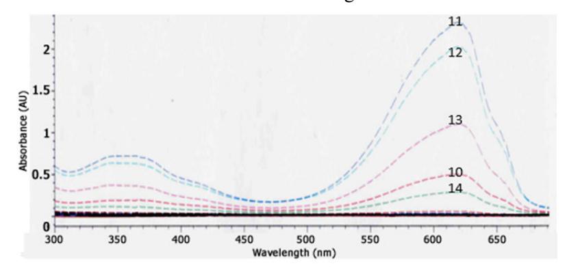
Figure 1 UV-Vis spectrum of gel chromatography fractions.
Gel filtration chromatography was performed using a Sephadex G-25 column of 25 x 2 cm and an elution flow rate of 0.5 mL/min. With this system, five blue fractions with different intensities were obtained, namely 10, 11, 12, 13 and 14, respectively. Based on the UV-Vis spectra, the five blue fractions were phycocyanin. Fraction 11 showed the highest purity ratio (1.730). The UV profile spectrum of all fractions is shown in Fig. 1. Thus, the purity of the phycocyanin increased from the extraction step by freeze thawing to the gel filtration chromatography step.
3.3 Characterization of Phycocyanin
Gel electrophoresis was performed on selected fractions of the gel filtration result (fractions 11-14) using SDS-PAGE (sodium dodecyl sulfatepolyacrylamide gel electrophoresis). Phycocyanin is composed of two different polypeptides based on their molecular weight, where the polypeptide with a low molecular weight is called unit α (BM: 12-19 kDa) and the polypeptide with a high molecular weight is called β unit (BM: 14-21 kDa) [15]. The electropherogram (Figure 2) shows that all fractions had a molecular weight of around 15-16 kDa. They were compared with a marker protein and phycocyanin (Sigma Aldrich).
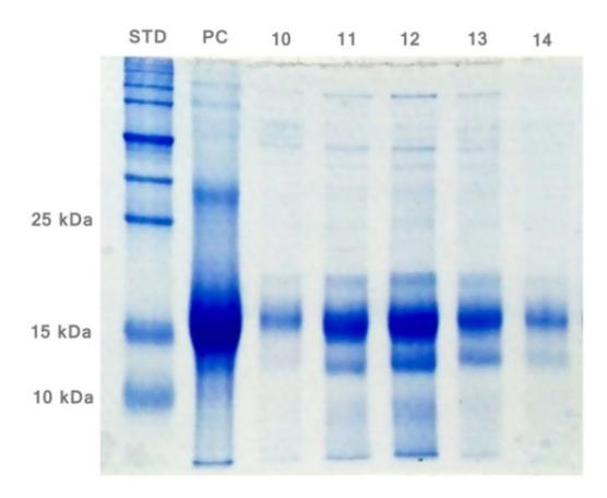
Figure 2 Electropherogram of gel filtration chromatography fractions (STD = protein marker; PC = phycocyanin (Sigma Aldrich); 10,11,12,13, and 14 = gel chromatography fractions).
Phycocyanin is a molecule containing an open tetrapyrrole chain. The tetrapyrrole chain is a chemical compound containing four pyrrole rings. A pyrrole ring consists of four carbon atoms and one nitrogen atom (C4H5N). In addition, phycocyanin has an amide group. The infrared spectra of the isolate showed the presence of amide groups at the 1646 cm1 wave number, corresponding to the area of the wave numbers for the amide group, i.e. 1690- 1630 cm1 (Sigma Aldrich). This amide group was used for the analysis of secondary proteins, which showed α-helix as the main element of the secondary structure [7]. In addition, bands in the 1000-1500 cm1 wave number area indicated the existence of a pyrrole ring [16]. The similarity of the infrared
spectrum pattern of the isolate (fraction 11) with standard phycocyanin in Fig. 3 confirmed that the isolated pigment was phycocyanin.
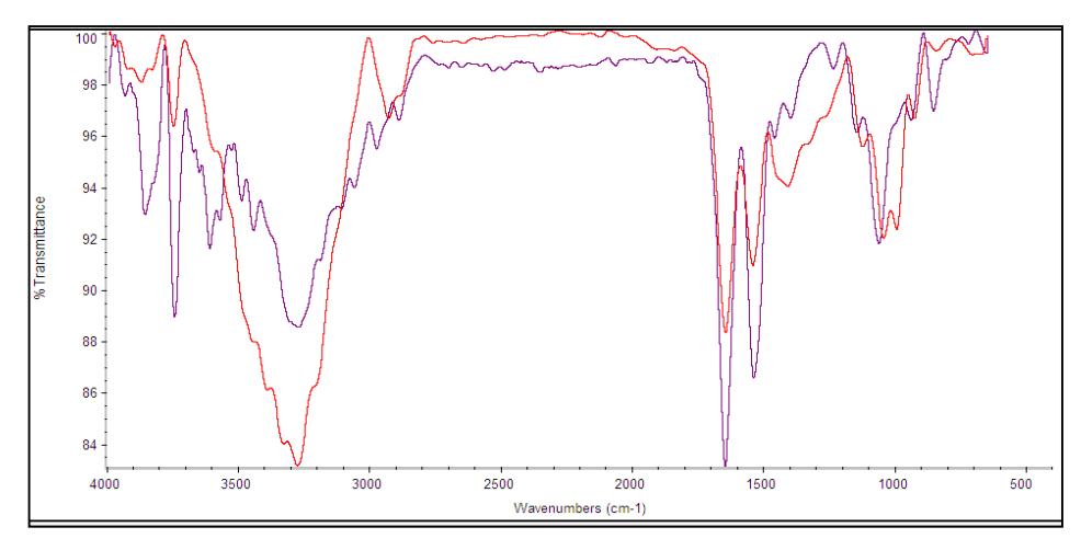
Figure 3 Overlay infrared spectrum of isolate (purple) and standard (red) phycocyanin.
4 Conclusions
Freezing-thawing with 0.01M phosphate buffer pH 7 was found to be the best extraction method of phycocyanin pigment since it shows higher C-PC content, purity ratio and yield than the other methods studied. The purity of the phycocyanin after purification by precipitation, dialysis and gel filtration was more than enough for food grade (≥ 0.7). Pigment characterization using SDS-PAGE showed a molecular weight of 15-16 kDa and its infrared spectrum confirmed it as phycocyanin.
Acknowledgment
This study was supported by Fundamental Research 2016 DIKTI, Directorate of Higher Education of the Ministry of Research, Technology and Higher Education of the Republic of Indonesia.