
1 Introduction
Indigenous people of Sumatra have used Sumatran plants since the beginning of civilization as foodstuff, aromatic agents, dyestuff, insecticidal agents, and medicine. Until the early 1960s, they used those medicinal plants for their primary health care; they only consulted a doctor in the nearest town when a traditional remedy could not cure their illness. Based on an inventory and ethnobotanical surveys carried out since 1982, more than 125 plants with traditional uses and barely touched scientifically have been selected for further chemical study [1].
Cancer is one of the leading causes of death. In Asia, the breast cancer rate is growing more rapidly compared to that in the West. As stated by the World Health Organization (WHO), nearly 1.38 million cases of breast cancer were diagnosed in 2008. The report also revealed that its prevalence reached 23% of all cancer incidences in the world. In reference to the WHO's statement, 209,000 new incidents have been found, particularly in Southeast Asia [2]. According to the International Agency on Research in Cancer, breast cancer has become the most common malignant tumor among Indonesian women [3]. Oral cancer, on the other hand, is one of the most often detected cancers in the world. In some South-Central Asian countries, the incidence of death caused by this cancer has become an important public health matter. Globally, this illness is normally detected after late medical diagnosis and leads to high mortality rates. Squamous cell carcinomas are the most common among oral malignancies [4]. Cases of oral cancer are estimated at about 275,000 for oral and 130,300 for pharyngeal cancers per year, not including nasopharynx. Two-thirds of these incidences occur in developed nations [5]. Different kinds of cancer therapies and complementary agents have been developed for their treatment.
A. denticulata, A. malaccensis, A. submutica, C. asiatia, C. zedoaria, M. indica, M. frondosa, and P. crocatum are among the Sumatran plants that have been used by Indonesian ethnic groups as a source of complementary and alternative medicine. These plants have been used for prevention and treatment of various cancer diseases. Some of these medicinal plants have recently attracted the attention of scientific researchers due to their accessibility, therapeutic effects and minimum side effects [6,7].
The aim of the present study was to obtain sufficient amounts of the major constituents of Stereocaulon halei Lamb (Stereocaulaceae) by isolation and to screen them for their cytotoxic activities. These isolated compounds, together with methanolic extracts of some Sumatran medicinal plants traditionally used as complementary agents for cancer treatment, i.e. rhizomes of A. denticulata, A. malaccensis, A. submutica, and C. zedoaria, aerial parts of C. asiatia, leaves of M. indica and P. crocatum, petals of M. frondosa and eight isolated compounds previously isolated from some Sumatran medicinal plants; crocatin A (2) and crocatin B (3) [8], curcumin (4) [9], demethoxymatteucinol (5) [10], mangiferin (7) [11], methyl caffeate (8) [12], tiliroside (10) [13] and usnic acid (11) [14], were tested for their cytotoxic activity towards MCF-7 human breast adenocarcinoma cell lines and HSC-3 human oral squamous cell carcinoma cell
lines by MTT assay (3-(4,5-dimethylthiazol-2-yl)-2,5-diphenyltetrazolium bromide) as described in [15], with a slight modification [16]. The structures of all isolated compounds (1-11) are shown in Figure 1.
\[\text{[rumus tidak dapat ditampilkan dengan baik — lihat PDF asli]}\]
Figure 1 Structures of isolated compounds of atranorin (1), crocatin A (2), crocatin B (3), curcumin (4), demethoxymatteucinol (5), lobaric acid (6), mangiferin (7), methyl caffeate (8), methyl-β-orcinol carboxylate (9), tiliroside (10), and usnic acid (11).
2 Materials and Methods
2.1 General Experimental Procedure
2.1.1 Chemical
The isolated compounds of the Sumatran medicinal plants crocatin A (2) and crocatin B (3) [8], curcumin (4) [9], demethoxymatteucinol (5) [10], mangiferin (7) [11], methyl caffeate (8) [12], tiliroside (10) [13], and usnic acid (11) [14] were available from previous work in our laboratory.
All organic solvents, i.e. n-hexane, ethyl acetate (EtOAc), methanol (MeOH), acetone, and formic acid were re-distilled, while the chloroform (CHCl<sub>3</sub>), toluene, and dimethyl sulfoxide (DMSO) were analytical grade. Chromatographic separation was performed using vacuum liquid chromatography on silica gel (Merck 35-70 \(\mu\)m) or radial chromatography on silica gel GF<sub>254</sub> (Merck). TLC analyses were performed on silica gel 60 F<sub>254</sub> (Merck). Visualization of TLC plates was performed using UV light at 254 and 365 nm and spraying with anisaldehyde-H<sub>2</sub>SO<sub>4</sub>, followed by heating.
Dulbecco's Modified Eagle Medium (DMEM), Penicillin-Streptomycin, TripLE Express<sup>TM</sup>, Fetal Bovine Serum (FBS), and 3-(4,5–dimethylthiazol-2-yl)-2,5-diphenyltetrazolium bromide (MTT) were obtained from Invitrogen Corporation (USA). Doxorubicin, cisplatin, and phosphate buffer saline (PBS) were purchased from Sigma Aldrich (USA).
MCF-7 and HSC-3 cell lines were kindly donated by Prof. Masa-Aki Ikeda of Tokyo Medical and Dental University. These cells were maintained in sterile complete growth media containing DMEM, supplemented with 10% of FBS and 1% of Penicillin-Streptomycin in 5% CO<sub>2</sub> at 37 °C.
2.1.2 Instruments
UV spectra were run on a Shimadzu Pharmaspec 1700. FT-IR spectra were obtained with a Perkin-Elmer 16 PC IR spectrometer. Melting points were determined with an Innotech Melting Point Apparatus. <sup>1</sup>H and <sup>13</sup>C NMR spectra were measured at 500 and 125 MHz on a Brucker Avance II 500 MHz NMR spectrometer and on a Bruker DMX 500 WB NMR spectrometer, at 400 and 100 MHz, on a Bruker Avance III 400 or at 300 and 75 MHz, on a Bruker 300 NMR spectrometer, respectively. Other equipments used were Dynatech MR5000 TECAN microplate reader (Tecan Group Ltd., Switzerland), Class II biological safety cabinet Jouan MSC 12 (Thermo Fischer Scientific, USA), CO2 Incubator NU-5100E (Nuaire, USA), Light microscope Motic B2 series (Motic Asia, Hongkong), Haemocytometer (Hirschmann, EM Techcolor,
Germany), Autoclave HV-85 (Hirayama, Japan), Centrifuge Universal 32 R (Hettich Zentrifugen, USA), Vortex mixer 2 x 3 Velp Scientifica.
Evaporation of organic solvents was carried out using a vacuum rotary evaporator (BUCHI Rotavapor R-210) at 60 °C and 300 mbar. Radial preparative chromatography was done using a Harrison Research Instruments Chromatotron®.
2.2 Plant Materials
Lichen S. halei Lamb was collected in January 2018 from rocks near the summit (at 2800 m) of Mount Singgalang (2877 m), West Sumatra, Indonesia. The plant specimens were identified by Dr. Friardi Ismed based on previous identification by Dr. Harrie Sipman (Berlin Museum). The voucher specimens were deposited at the Herbarium of Andalas University (ANDA), West Sumatra (Indonesia) with reference numbers JB/09/117 and GSb 1 respectively.
Plant samples were collected around the city of Padang in November 2017 and identified by Dr. Nurainas from Herbarium of Andalas University (ANDA) by direct comparison to samples from the collection of herbarium specimens available at the Herbarium. A. denticulata, A. malaccensis and A. submutica were compared to the specimens collected by Suci Erta Fitri, et al. from the Department of Biology, Faculty of Mathematics and Natural Science, Andalas University, with reference numbers 01 – Sc Al HR (ANDA, fl, fr.), 03 AL SC SBK, and 04 – Sc AL HR (ANDA, fl.) respectively. C. zedoaria was compared to the specimen collected by R. Tamin with reference number 2138. C. asiatica was compared to the specimen collected by Sri Suhartini with reference number 01. M. indica (coll. number 327), M. frondosa (coll. number 327) and P. crocatum (Red Betle, coll. number 198) were compared to specimens of those plants that have previously been identified at the Herbarium.
2.3 Extraction and Isolation
Fresh rhizomes of A. denticulata, A. malaccensis, A. submutica, and C. zedoaria, aerial parts of C. asiatia, leaves of M. indica and P. crocatum, and petals of M. frondosa (100 g each) were chopped into small pieces and macerated with MeOH overnight at room temperature. This process was repeated thrice, after which each combined extract was filtered through filter paper and evaporated in vacuo to get thick extracts of rhizomes of A. denticulata (532 mg), A. malaccensis (365 mg), A. submutica (129 mg), and C. zedoaria (3.35 g), aerial parts of C. asiatia (4.54 g), leaves of M. indica (12.52 g) and P. crocatum (5.33 g), petals of M. frondosa (11.36 g), respectively which were stored at 4 °C in a refrigerator for future chemical and biological analysis.
Dried material thalli of S. halei Lamb (387 g) were extracted in turn with n-hexane, EtOAc, and acetone (3 times x 1 L). During this extraction process contact with the alcoholic solvents was entirely avoided. Evaporation of n-hexane solvent in vacuo gave a yellowish solid that showed one major spot on TLC, which after recrystallization from CHCl<sub>3</sub>-n-hexane gave colorless needles of atranorin (1, 83 mg).
Attempts to isolate minor components from this n-hexane extract were not successful due to the limited amount of sample available. After flash chromatography on silica gel, the EtOAc extract was separated by radial chromatography on the same adsorbent, eluted with an increasing amount of CHCl<sub>3</sub> in n-hexane to give atranorin (1, 479 mg) and methyl-\(\beta\)-orcinol carboxylate (9, 19 mg).
After evaporation of the solvent, the acetone extract gave a solid residue (2.50 g), which was separated by radial chromatography on silica gel using the Chromatotron<sup>®</sup>, eluted with an increasing amount of CHCl<sub>3</sub> in the n-hexane to give fractions. Most nonpolar fractions that showed the same pattern on the TLC plates were merged, evaporated, and recrystallized from the CHCl<sub>3</sub>-n-hexane to give atranorin (1, 114 mg). Mother liquor and the rest of the fractions were combined, evaporated, radial chromatographed on silica gel and then eluted with toluene-EtOAc-formic acid (80:15:5) to give fractions. The fractions with the same patterns on TLC plates were combined, evaporated and recrystallized from the CHCl<sub>3</sub>-n-hexane to give atranorin (1, 10 mg), lobaric acid (6, 11 mg), and methyl-β-orcinol carboxylate (9, 5 mg).
Atranorin (1); colorless needles; mp 190-191 °C (undepressed in admixture with reference compound [17]); IR \(\nu\)max (cm<sup>-1</sup>) = 2958, 2014, 1640; UV (MeOH) \(\lambda_{max}\) (log ε): 269 nm (6.99), 325 nm (7.08); <sup>1</sup>H NMR (500 MHz, DMSO) δH 2.05 (3H, s, Me-8'), 2.36 (3H, s, Me-9'), 2.41(3H, s, Me-9), 3.98 (3H, s, CO2Me), 6.42 (1H, s, H-5'), 6.51 (1H, s, H-5) and 10.22 (1H, s, -CHO); <sup>13</sup>C NMR (125 MHz, CDCl<sub>3</sub>): δC 102.92 (C-1), 196.17 (C-2), 108,63 (C-3), 167.57 (C-4), 112.93 (C-5), 152.51 (C-6), 169.77 (C-7), 193.92 (C-8), 25.67 (C-9), 116.86 (C-1'), 162.95 (C-2'), 110.34 (C-3'), 152.06 (C-4'), 116.09 (C-5'), 139.94 (C-6'), 172.27 (C-7'), 24.12 (C-8'), 9.46 (C-9'); Rf 0.86 with eluent G (toluene-EtOAc-formic acid (70:25:5).
Methyl-β-orcinol carboxylate (9); colorless needles; mp 140-141 °C undepressed in admixture with reference compound [17]); IR \(\nu\)max (cm<sup>-1</sup>) = 3746, 3395, 3002, 2881, 1993, 1802, 1616; UV (MeOH) \(\lambda_{max}\) (log ε): 267 nm (6.65), 304 nm (6.7); <sup>1</sup>H NMR (500 MHz, DMSO) δH 1.95 (3H, s, 9-Me), 2.36 (3H, s, 8-Me), 3.85 (3H, s, COOMe), 6.28 (1H, s, H-5), 11.56 (1H, bs, 2-OH); <sup>13</sup>C NMR (125 MHz, DMSO-\(d_6\)) δC 104.04 (C-1), 159.96 (C-2), 108.11 (C-3), 161.6 (C-4), 110.48 (C-5), 138.68 (C-6), 171.77(C-7), 7.95 (C-8), 23.39 (C-9), 51.88 (COOMe-10).
Lobaric acid (6); colorless needles; mp 196-197 °C (undepressed in admixture with reference compound [17]); IR νmax (cm-1) = 3744, 3150, 2956.11, 2872, 2771, 2152, 1979, 1683, 1558; UV (MeOH) λmax (log ε): 210 nm (6.98), 266 nm (7.08), 296 nm (7.13); 1 H NMR (500 MHz, acetone-d6) δH 0.95 (3H, m, 5'''), 1.43 (4H, m, 4'''), 1.55 (2H, m, 3'''), 1.68 (2H, m, 2'''), 2.88 (2H, t, J=7.25), 3,29 (2H, t, J=1.00, 1'''), 3.99 (3H, s, OCH3-4), 6.79 (1H, s, 3'), 7.03 (1H, dd, J=2.4, 2.5, 3,5); 13C NMR (125 MHz, acetone-d6) δC 111.64 (C-1), 162.83 (C-2), 106.04 (C-3), 164.58 (C-4), 111.33 (C-5), 149.32 (C-6), 161.45 (C-7), 202.38 (C-1''), 41.34 (C-2''), 25.79 (C-3''), 22.29 (C-4''), 13.30 (C-5''), 111.35 (C-1'), 159.92 (C-2'), 107.12 (C-3'), 148.29 (C-4'), 138.96 (C-6'), 171.18 (C-7'), 27.79 (C-1'''), 31.00 (C-2'''), 32.00 (C-3'''), 21.83 (C-4'''), 13.45 (C-5'''), 56.01 (OCH3-4); Rf 0.44 with eluent G (toluene–EtOAc–formic acid (70:25:5).
2.4 MTT Assay
Cytotoxic activity was measured by MTT assay following the protocol developed by Mosmann [15], with a slight modification [16]. The extracts and isolated compounds were dissolved in DMSO and serially diluted in DMEM to obtain different concentrations (3.125, 6.25, 12.5, 25, 50 and 100 µg/mL). In 96 well plates, 100 µL of suspension cells with density of 2 x 10⁵ cells/mL were added and incubated at 37 °C in a 5% (v/v) CO2 atmosphere for 24 h. Next, the cells were treated with various concentrations of extracts, isolated compounds, and positive control in triplicate and incubated for 24 hours.
After incubation, the culture medium was removed and then the cells were washed with PBS once. After addition of 20 µL of MTT solution at a concentration of 5 mg/mL in PBS to each well, the medium was then incubated for 4 hours, the supernatant was discarded and the insoluble formazan crystals were diluted in 100 µL of DMSO. Finally, it was left in a dark place at room temperature for an additional 1 h.
The absorbance of the formazan solution was determined at 570 nm with reference at 630 nm using a microplate reader. Untreated cells were used as negative control while DMEM complete media was used as solvent control. Doxorubicin was used as positive control for the MCF-7 cell line and cisplatin was taken as positive control for the HSC-3 cell line. The cell viability (%) was counted as the relative percentage of the average absorbance of the treated cells compared to the untreated control cells.
The IC50, or the half maximal inhibitory concentration, was measured as the concentration of the extract and compound that causes 50% inhibition in cell viability towards the MCF-7 and HSC-3 cell lines. Cell viability (%) was calculated as follows:
Cell viability (%) = \[\frac{(A_{samples} - A_{blank})}{(A_{untreated} - A_{blank})} x100\]
The IC50 values were defined by plotting graphs of cell viability versus concentration.
2.5 Statistical Analysis
The statistical analysis of the experimental data was done by comparing the same treatment of the samples as a group with the untreated cells (negative control) and represented in the form of means ± standard error. The statistical significance of the data was computed using one-way analysis of variance (ANOVA; 95% confidence interval), along with a Dunnet post hoc test for the determination of the level of significance. The results are indicated as significant if P < 0.05.
3 Results and Discussion
3.1 Results
Out of 387 g dried-weight S. halei Lamb, three compounds, i.e. atranorin (1, 686 mg), lobaric acid (6, 11 mg), and methyl-β-orcinol carboxylate (9, 24 mg), were isolated. Attemps to isolate lobarine, which could be detected on the TLC, were not successful due to the limited amount of sample available.
Tabel 1 IC50 Values of methanolic extracts of Sumatran medicinal plants on MCF-7 cell lines.
| Groups | Plant species | IC50 (µg/mL) |
|---|---|---|
| ADM | A. denticulata | 1,176.63 |
| AMM | A. malaccensis | 309.53 |
| ASM | A. submutica | 70.95 |
| CAM | C. asiatica | 615.70 |
| CZM | C. zedoaria | 222.54 |
| MIM | M. indica | 4,615.38 |
| MFM | M. frondosa | 177.14 |
| PCM | P. crocatum | 57,626.25 |
| Positive control | Doxorubicin | 3.38 |
Table 1 shows the IC50 values of each methanolic extract of rhizomes of A. denticulata, A. malaccensis, A. submutica, and C. zedoaria, aerial parts of C. asiatia, leaves of M. indica and P. crocatum, petals of M. frondosa against
MCF-7 cell lines. Most extracts tested did not show significant activities against the MCF-7 cell line, except A. submutica and M. frondosa, which showed weak cytotoxic activity against MCF-7, as shown in Figure 2.
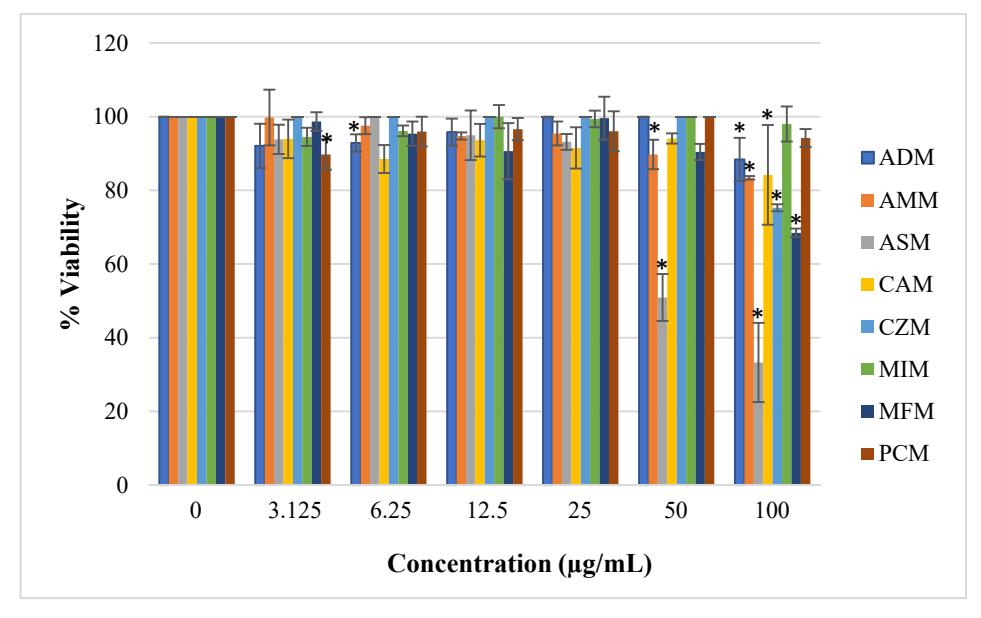
Figure 2 Effects of different concentrations of methanolic extracts of Sumatran medicinal plants on percentage of MCF-7 cell viability. Doxorubicin with an IC<sub>50</sub> value of 6.21 μM was taken as positive control. The results represent the mean \(\pm\) SEM of triplicate experiments. *(P < 0.05) means a significant difference with control.
Table 2 IC<sub>50</sub> value of compounds 1-11 on MCF-7 and HSC-3 cell lines.
| Cuouna | Companyed and mathematic sytmet | \(IC_{50}(\mu M)\) | |
|---|---|---|---|
| Groups | Compounds and methanolic extract | MCF-7 | HSC-3 |
| 1 | atranorin | 208.20 | 474.08 |
| 2 | crocatin A | 331.22 | 283.02 |
| 3 | crocatin B | n.d | 434.17 |
| 4 | curcumin | 12.06 | 208.72 |
| 5 | demethoxymatteucinol | 383.03 | n.d. |
| 6 | lobaric acid | 172.05 | 88.92 |
| 7 | mangiferin | 1,112.01 | n.d. |
| 8 | methyl caffeate | n.d | 100.37 |
| 9 | methyl-\(\beta\)-orcinol carboxylate | 382.60 | 260.09 |
| 10 | tiliroside | n.d | 41.06 |
| 11 | usnic acid | 255.87 | 44.32 |
| AMM | A. malaccensis | 294.82a | n.d. |
| Control | Doxorubicin | 6.21 | - |
| Cisplatin | - | 20 | |
Note: n.d. = concentration in \(\mu\)g/mL not determined; .a = concentration in \(\mu\)g/mL.
A summary of the cytotoxic activities of compounds 1-11 is presented in Table 2. The effects of the various concentrations of compounds 1-11 and the extracts of Sumatran medicinal plants on the percentage of MCF-7 and HSC-3 cell viability are shown in Figures 3 and 4, respectively.
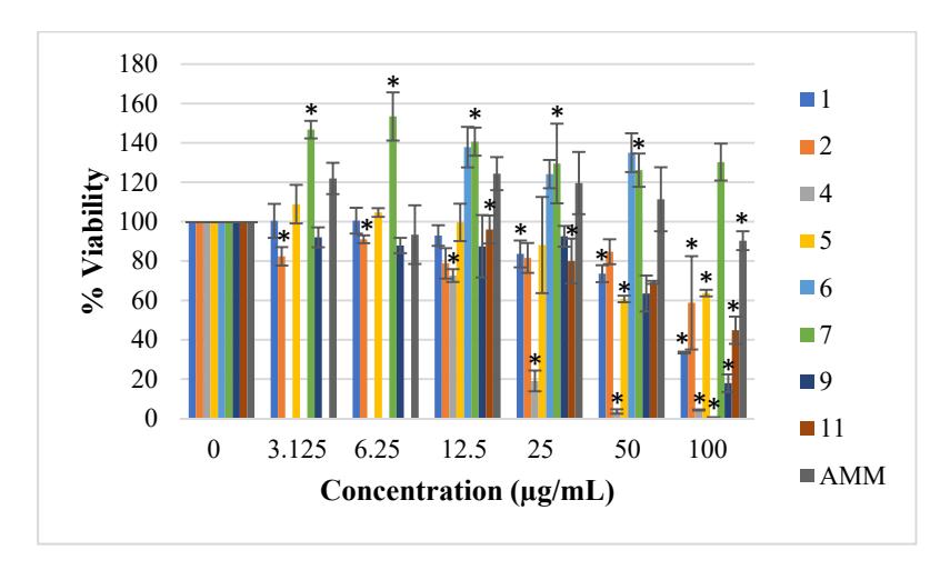
Figure 3 Effects of various concentrations of compounds 1-11 and extracts of Sumatran medicinal plants on percentage of MCF-7 cell viability. Doxorubicin with an IC<sub>50</sub> value of 6.21 \(\mu\)M was taken as positive control. The results represent the mean \(\pm\) SEM of triplicate experiments. *(P < 0.05) means a significant difference with control.
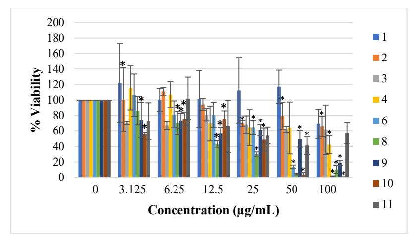
Figure 4 Effects of various concentrations of isolated compounds of Sumatran plants on percentage of HSC-3 cell viability. Cisplatin was with an IC<sub>50</sub> value of 20 \(\mu\)M used as positive control. The results represent the mean \(\pm\) SEM of triplicate experiments. *(P < 0.05) means a significant difference with control.
3.2 Discussion
Air-dried thalli of S. halei Lamb were extracted in turn with n-hexane, EtOAc, and acetone. Based on experience from our previous work special attention was taken to avoid contact with alcoholic solvents to prevent trans-esterification. Due to the moderate success rate of cancer treatment with modern medicines, many cancer patients in Indonesia still use traditional medicinal plants as complementary treatment. Some extracts of Sumatran medicinal plants have been investigated for their cytotoxicity.
C. zedoaria, used in jamu (Indonesian traditional medicine), is commonly consumed for the medication of breast and cervical cancer [18]. In a previous study, water and ethanolic extracts of C. zedoaria have been examined for cytotoxic activity against large-cell lung carcinoma (CORL-23) and prostate cancer (PC3) and one type of normal human cell line (fibroblast cells (10FS). The cytotoxic activity of ethanolic extracts of C. zedoaria towards large-cell lung carcinoma (CORL-23), prostate cancer (PC3) and one type of normal human cell line (fibroblast cells (10FS) was examined. The ethanolic extracts of C. zedoaria exhibited a cytotoxic effect towards CORL-23, PC3 and 10FS was found to be less toxic against 10FS (IC50: 6.05, 17.84 and 55.50 µg/mL, respectively). The water extract of this plant did not exhibit cytotoxicity towards all of the human cells used in the experiment, whereas the ethanolic extract did show cytotoxic activity towards lung cancer cells but was less active against normal cells [19].
The antimetastatic activity of C. zedoaria Roscoe (CZ) on pulmonary metastasis of melanoma cells (B16) has been examined. The input of CZ (250 and 500 mg/kg) for 6 weeks from 2 weeks before tumor inoculation considerably reduced the number of metastatic surface nodules in the lung as well as a prolonged survival rate [20]. In the present work, the methanolic extract exhibited weak activity towards MCF-7 cells.
A previous study found that cytotoxicity of hexane, dichloromethane, ethyl acetate, and methanolic extract of C. zedoaria rhizomes on MCF-7 was 18.4 ± 1.6 µg/mL, 40.6 ±2.3 µg/mL, > 100 µg/mL, and > 100 µg/mL, respectively, and towards cervical cancer (Ca Ski) cell lines it was 19.0 ± 1.5 µg/mL, 83.5 ± 2.7 µg/mL, > 100 µg/mL, and > 100 µg/mL, respectively [21]. We found in our experiment that the IC50 value of methanolic extract of C. zedoaria towards the MCF-7 cell line was 222.54 µg/mL. Both the IC50 values of methanolic extract from the previous study and our study gave weak inhibitory activities towards the MCF-7 cell line.
Curcuma rhizome with its main constituent curcumin (4) has been widely studied for its anti-cancer activity based on its use in Indian and Chinese remedies. The IC50 values of curcumin (4) towards MCF7 cell lines was calculated in the prior study at an IC50 value of 79,58 µg/mL, whereas the IC50 value of curcumin (4) was not determined in our study. It has been proved that curcumin convincingly inhibited the growth of MCF-7 cells as indicated by induced apoptosis in a dose- and time-dependent manner, followed by a decrease of MCF-7 cell viability [22].
In Indonesia, leaves of P. crocatum are used for various deseases, including cancer. Wicaksono, et al. [23] have shown that methanolic extract of P. crocatum exhibits cytotoxic effect against human breast cancer (T47D) cell lines with an IC50 value of 44.25 µg/mL by inhibiting p44/p42 phosphorylation. Our work showed that the extract of the leaves of this plant was inactive against the MCF-7 cell line and its main constituents, crocatin A (2) and crocatin B (3), exhibited weak activity.
It has been documented that atranorin (1), lobaric acid (6), and usnic acid (11) exhibit cytotoxicity on MCF-7 [24,25]. Bačkorová, et al. [24] have reported a higher IC50 for atranorin (1) and usnic acid (11) than in this study, which could be explained by the difference in experimental conditions, such as time (72 vs 24 hours in our study), growth medium used (RPMI 1640 vs DMEM in our study), combination of antibiotics in the medium (100 U/mL of penicillin, 100 µg/mL of streptomycin and amphotericin 25 µg/mL vs 1% Penicillin-Streptomycin in our study). For lobaric acid (6), the isolated compounds of lichen exhibited weaker activity against MCF-7 cells in both our study and a previous experiment [24] with an IC50 of 172.05 µM and >100 µM, respectively.
Demethoxymatteucinol (5) was isolated from Sumatran medicinal plant Sphaerostephanos polycarpus, which is traditionally used as an anti-infection agent [10]. It showed weak activity towards the MCF-7 cell line.
In the previous in-vitro cytotoxicity study on human oral epidermoid carcinoma cells, leaf extract of Guazuma tomentosa was evaluated for malignant melanoma, lung carcinoma, ileocecal adenocarcinoma, epidermoid carcinoma, malignant melanoma, and also medulloblastoma cells [26]. Tiliroside (10) as a major constituent of G. tomentosa was previously examined for its in-vitro cytotoxic activity towards the following T-cell lines: CCRF CEM, H33AJ-JA13, HUT 78 and H9, B cell lines (NAMALWA, JIYOYE, CCRF-SB), proerythrocytes (K562), monocytes (U937). It showed a cytotoxic effect towards two of the tested nine cancer cells, CCRF-CEM and NAMALWA, with an IC50 value of 17.1 and 16.1 µg/mL, respectively [27].
Tiliroside (10) also showed cytotoxic activity towards two human hormonedependent breast cancer cell lines (T47D and MCF7) with an IC50 of 67.79 and 112.77 µg/mL, respectively [28], whereas the IC50 of tiliroside (10) against MCF-7 in our study could not be identified.
Methyl caffeate (8) was isolated from Balanophora elongata Blume, which is traditionally used as an anti-infection agent [12]. The IC50 of this compound was not determined but it has been reported as being active against MCF-7 with an IC50 value of 0.62 µM in [29]. Our findings showed that this compound had weak activity against the HSC-3 cell lines.
Previous studies have reported that mango leaf extract and mangiferin (7) exhibited cytotoxic activities against ductal carcinoma (BT474), bronchogenic carcinoma (Chago-K1), liver hepablastoma (Hep-G2), gastric carcinoma (Kato-III), colon carcinoma (SW620) cells with IC50 values of > 200 µg/mL, for all cancer cells [30]. Kernel extract of M. indica was tested for cytotoxic activity towards MCF-7 and MDA-MB-231 cells and revealed a cytotoxic effect with an IC50 value of 30 and 15 μg/mL, respectively [31].
Mangiferin (7) showed the ability to re-sensitize MCF-7 cell lines treated with short-term doxorubicin in-vitro by modulating efflux transporters, Pglycoprotein (P-gp), MRP1 and BCRP. Mangiferin (7) at high concentrations is considered to act as a chemosensitizer for chemotherapy using doxorubicin [32]. However, in this study, methanolic extract of M. indica and mangiferin (7) did not show cytotoxic activity (IC50 values > 1000 µM).
The indication of induced apoptosis of methanolic extracts of C. asiatica towards MCF-7 was demonstrated by nuclear condensation, increased annexin staining, loss of mitochondrial membrane potential, and induction of DNA breaks indicated by TUNEL reactivity [33]. In the present study, the in-vitro antiproliferative effect of the methanolic extract of the aerial parts of C. asiatica towards MCF-7 cancer cells was observed to be weak (IC50 of 615.70 µg/mL).
In indigenous usage, the whole plant of M. frondosa is boiled and its decoction is given to treat fever, asthma and coughing [34]. M. frondosa has various pharmacological activities, such as antiseptic, antioxidant, antidermatitic, fungicide, insecticide and antitumor activities [35]. It has also been found to possess antibacterial effects [36]. Our study showed that the methanolic extract of M. frondosa had weak cytotoxic activity against MCF-7 (IC50 value of 177.14 µg/mL).
4 Conclusions
The present study demonstrated that the methanolic extract of rhizomes of A. submutica, with an IC50 value of 70.95 µg/mL, is the most active towards breast adenocarcinoma (MCF-7) cell line compared to the methanolic extracts from the other plants tested in this study. It also showed that all of the isolated compounds tested had weak or unsignificant activity against MCF-7 and HSC-3 cell lines, except atranorin (1), lobaric acid (6) and methyl-β-orcinol carboxylate (9), with activity towards MCF-7 208.20 µM, 172.05 µM, and 382.60 µM, respectively. Meanwhile, the IC50 value of lobaric acid (6) and methyl-β-orcinol carboxylate (9) against the HSC-3 cell line was obtained at 260.09 µM and 88.92 µM, respectively. None of the extracts tested, except for the methanolic extract of A. submutica, showed significant activity towards MCF-7. Future research is still needed to identify and purify the active chemical constituents of this species for their cytotoxic agents.
The IC50 value of lobaric acid (6) indicated that it has the most cytotoxic activity towards MCF-7, followed by atranorin (1) and methyl-β-orcinol carboxylate (9). Towards the HSC-3 cell line, lobaric acid (6) and methyl-βorcinol carboxylate (9) were found to have moderate cytotoxic effects. However, our results indicated that atranorin (1), lobaric acid (6), and methyl-βorcinol carboxylate (9) as a member of depside and its derivative depsidone were widely distributed among the lichens, can be seen as promising lead compounds for human breast adenocarcinoma and human oral squamous cell carcinoma cancer drugs. Hence, the structure-activity relationships (SARs) of atranorin (1), methyl-β-orcinol carboxylate (6), and lobaric acid (9) and their derivatives are worthy of investigation.
Acknowledgements
The authors gratefully acknowledge the research grant of the Ministry Research, Technology and Higher Education of the Republic of Indonesia received by DA under the DP2M-DIKTI Competence Grant 2015-2017. We thank the Ministry of Health of the Republic of Indonesia for the use of the Indonesian Herbal Pharmacopoeia markers from the BBO and BBOT projects, Dr. Nurainas from the Herbarium of Andalas University (ANDA) for the identification of the plant specimens, and Professor Hamburger from the University of Basel, Switzerland for the NMR spectra of compounds 1, 6 and 9.