
1 Introduction
Micromelum minutum (G. Forst.) Wight & Arn. is a vegetation found in Indonesia. This particular plant is not widely known, although it has been widely used since ancient times in several nations as a traditional medicine to treat fever, dizziness, and dyspepsia [1][2]. The available data on this plant in Indonesia is only limited to the distribution area and there is no phytochemical data or data on its pharmacological activity.
Isolation and identification of active compounds in M. minutum leaves from several countries has been done. M. minutum grown in Malaysia [3]-[5], Thailand [6]-[8], India [7] and Sri Lanka [9] contains coumarin as a main compound. Additionally, other compounds, such as flavonoids and alkaloids, which are known to be active as anticancer agents, are also found in this plant [4],[10],[11]. However, there are variations in the structure of these compounds depending on the location of the plant. Although in some countries the isolation and identification of compound contents has been carried out, M. minutum grown in Indonesia, also known as Lada-lada in Sulawesi [12], has not yet been explored.
Cancer is a global health problem and is the second leading cause of death in the world. A type of cancer that occurs in many women is breast cancer. The most common cancer subtypes among Indonesian women aged 40 to 44 years are luminal type A and triple negative [13][14]. Chemotherapy does not only destroy cancer cells but also attacks normal cells [13]. Therefore, it is necessary to look for new drugs, especially from natural products, that can be used as a single therapy or in combination with chemotherapy [11],[15] to reduce toxicity and chemotherapy resistance that already exists.
Micromelum minutum is a potential resource for anticancer drugs from natural products. Cytotoxicity activities of Malaysian M. minutum extracts as well as active compounds have been proved on CEM-SS leukemic cells [5] and breast cancer cells [11]. Active compounds or extracts of Thai M. minutum exhibited cytotoxic activity toward leukemic cell lines [6] and lung, colon, and breast cell lines [8], respectively. Nevertheless, an investigation into the cytotoxicity of M. minutum grown in Indonesia has never been done. This study was carried out to examine the cytotoxic activity of Indonesian M. minutum leave extracts toward breast cancer cell lines as the initial step. Following the result, the isolation and identification of active compounds from the potential active extracts was carried out.
2 Materials and Methods
2.1 Extraction of Micromelum minutum Leaves
Fourteen kilograms of M. minutum leaves were collected from Bantimurung Bulusaraung National Park, Makassar, South Sulawesi, Indonesia in March 2017. The leaves were dried and determined at the Department of Pharmaceutical Biology, Faculty of Pharmacy, Universitas Gadjah Mada (UGM), Yogyakarta, Indonesia. The dried leaves (5.5 kg) were macerated gradually in hexane (HEM), ethyl acetate (EEM), and methanol (MEM) (technical grade, General Labora, Yogyakarta, Indonesia). Following
maceration, evaporation by using rotary vacuum evaporator (Büchi) was carried out to obtain crude extracts. The crude extracts were weighed to determine the extraction yield (%).
2.2 Chromatography Profiling of M. minutum Extracts
To obtain the content profile of the extracts, thin-layer chromatography (TLC) in the normal system was carried out. An aluminum TLC plate (105554, Merck) and chloroform (pro analysis, Merck) as mobile phase were used. The TLC spots were viewed under 254 nm and 366 nm UV light and under visible light after spraying with CeSO4 reagent (Merck).
2.3 Cytotoxic Assay
All breast cancer cell lines came from the collection of the Cancer Chemoprevention Research Center (CCRC), Faculty of Pharmacy UGM. The MCF-7 and T47D cells originally came from the American Type Culture Collection and were donated by Prof. Masashi Kawaichi, Department of Biological Sciences, Nara Institute of Science and Technology. The cells were cultured in DMEM (Dulbecco's Modified Eagle's Medium) complete media (Gibco) and cultured at 37 °C with 5% CO2atmosphere. Complete media were supplemented with antibiotics (150 µg/mL streptomycin and 150 U/mL penicillin) (Gibco, Invitrogen, USA), 1.25 µg/mL fungizone (Amphotericin B) (Gibco), and fetal bovine serum (FBS) (Sigma-Aldrich, USA) as much as 20% for MCF-7 cells [16] and 10% for 4T1 cells [17].
A cytotoxic assay was carried out by MTT method as previously modified for MCF-7 [16] and 4T1 [17] cells. The cells were distributed into a 96-well plate (1x10⁴ or 2.3 x 10³ cells/well for MCF-7 or 4T1 cells, respectively) and incubated in a 5% CO2 incubator (Heraeus) at 37 °C for 24 h. The medium was removed, the cells were rinsed with phosphate-buffered saline (PBS), and extract solutions in various concentrations were added in triplicate. The extracts were first diluted in dimethyl sulfoxide (DMSO (Merck) and then serially diluted (1, 5, 10, 25, 50, 62.5, 100, 125, 150, 200, 250, and 375 µg/mL). The final concentration of DMSO was not more than 0.6% for each concentration. This highest concentration of DMSO was employed as solvent control. After 24 h of incubation, the culture media were removed. Following PBS washing, 100 μL of 3-(4,5-dimethylthiazolul-2)-2,5-diphenyltetrazolium (MTT) (Sigma) solution at a concentration of 0.5 mg/mL was added to each well. The plate was incubated for 3 h until formazan crystals were formed. One hundred microliters of 10% sodium dodecyl sulfate in 0.01 N HCl as stopper solution was added to dissolve the formazan, followed by overnight incubation protected from light at room temperature. Then absorbance was measured using an ELISA reader at 595 nm. Control and blank samples were prepared as previously described in [15].
2.4 Data Analysis
The percentage of cell viability was determined and used to calculate the IC50 value, which is the concentration that inhibits 50% of cell viability, as previously described in [15].
3 Results
3.1 Chromatogram Profile of Micromelum minutum Extracts
Gradual maceration generated hexane extract (HEM), ethyl acetate (EEM), and methanol extract (MEM). Before evaporation, HEM was liquid, while EEM and MEM were semi-solid. After evaporation, the extracts were weighed and the yield was calculated, as presented in Table 1. The chromatogram profiles of each extract are shown in Fig. 1. By using the normal system, it was confirmed that HEM was dominated by non-polar compounds, MEM contained semi-polar and polar compounds, while EEM consisted of a more complex mixture of compounds, which ranged from non-polar to polar compounds.
Table 1 Yields of Indonesian Micromelum minutum extraction.
| Extract | Yield | |
|---|---|---|
| gram | % wt/dry wt | |
| Hexane (HEM) | 144.95 | 2.65 |
| Ethyl acetate (EEM) 336.76 | 6.12 | |
| Methanol (MEM) | 356.95 | 6.49 |
3.2 Cytotoxic Activities of M. minutum Extracts on Breast Cancer Cells
The Indonesian M. minutum leave extracts showed dose-dependent cytotoxic activity against MCF-7 and 4T1 breast cancer cells (Fig. 2). The detailed data can be found in the Supplementary Data and as part of the theses [18] and [19]. In general, the curve of HEM shifted to the left, indicating that it possessed the most potent cytotoxicity. MEM was the least potent of both cell lines. Calculation of the IC50 values confirmed that HEM had the strongest cytotoxicity, while MEM had the weakest cytotoxicity (Table 2). On MCF-7 cells, MEM at the highest dose did not decrease cell viability up to 50%, thus the calculation was based on extrapolation. Because of its low cytotoxicity, the IC50 value on 4T1 cells could not be determined. As comparison, the IC50 value of anticancer agent doxorubicin is 1.56 µg/mL in MCF-7 cells.
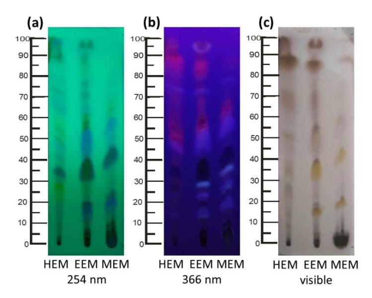
Figure 1 The thin-layer chromatogram profiles of Indonesian Micromelum minutum extracts. Normal-phase chromatography was employed, and the plate was viewed under UV 254 nm (a), UV 366 nm (b), and visible light after CeSO4 spray application (c).
Table 2 IC50 Values of Indonesian M. minutum Extracts on Breast Cancer Cells.
| IC50 (µg/mL) | ||
|---|---|---|
| 87 | ||
| 170 | ||
| 384* | nd | |
| MCF-7 cells 4T1 cells 148 185 | ||
* = extrapolated; nd = not determined
4 Discussion
This research is the first study on the chemical constituents and biological activity of Micromelum minutum leaves from Indonesia, which for M. minutum from several other countries have already been reported [3]-[11]. The extraction of Malaysian M. minutum leaves yielded nonpolar, semipolar, and polar extracts of 2.29, 5.72, and 7.25% w/w, respectively [4]. Extraction of Indonesian M. minutum leaves resulted in higher yields of nonpolar and semipolar extracts, but the polar extract yield was smaller than from Malaysian leaves (Table 1). These results indicate that less compounds can be extracted with polar solvent from Indonesian M. minutum than from Malaysian M. minutum.
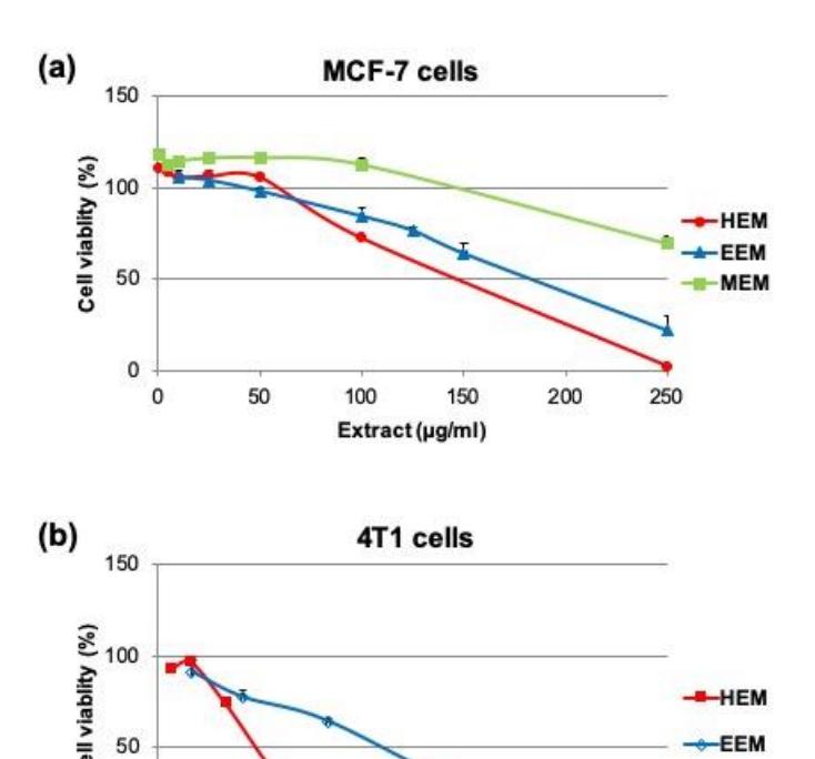
Figure 2 Effects of Indonesian M. minutum extracts on cell viability of breast cancer cells, MCF-7 (a) and 4T1 (b). The cells were treated with a serial concentration of hexane (HEM), ethyl acetate (EEM), or methanol extract (MEM) and incubated for 24 h. Then they were assayed by MTT method in triplicate. Except for the EEM on MCF-7 cells, the graph represents mean SE. As for EEM in MCF-7, the experiments were performed three times and the graph represents the mean SD. Detailed data for each experiment are described in the Supplementary Data.
Based on the biological activity study, it was confirmed that the extract with the most potent cytotoxicity was HEM, a nonpolar extract, followed by EEM, a semipolar extract (Fig. 2). An extract is considered to have a strong cytotoxic effect, with an IC50value less than 30 µg/mL [5]. This is the first study on cytotoxic activities of a nonpolar M. minutum extract toward breast cancer cells. Both HEM and EEM had similarly low potency on the MCF-7 cell line (a positive estrogen receptor) and the progesterone receptor cell line. On the other hand, HEM exhibited higher toxicity on 4T1, a triple negative breast cancer cell line. In accordance with a previous study, like Malaysian M. minutum methanol
extract is not potent on CEM-SS leukemic cells [5], methanolic extract from Indonesian M. minutum is not cytotoxic to breast cancer cell lines. It is plausible that active compounds responsible for the biological activity in the nonpolar extract are dominated by volatile oils [7],[9]. Our group studied the active compounds of the HEM nonpolar extract. The structures were successfully elucidated and the result was submitted for publication.
5 Conclusion
Hexane extract from Indonesian M. minutum leaves has the potential to be further developed as an anticancer agent, especially to treat triple negative breast cancer cells.
Acknowledgements
This research was funded by the Ministry of Research, Technology, and Higher Education, Indonesia through a Decentralized Research Grant 2017 given to RAS. We wish to express our gratitude to Mr. Heri Suryanto and Mr. Abdul Qudus, Makassar Center for Environment and Forestry Research and Development, South Sulawesi, Ministry of Environment and Forestry, Indonesia for the plant material collection. The authors state their contributions as follows: RAS and EM conceptualized and supervised the study; N and NAS performed the experiments; MI and N wrote the original paper and prepared the figures. All authors discussed the results and contributed to the final manuscript.