
1 Introduction
Cancer is a significant health problem worldwide. According to data from the International Agency for Research on Cancer, there were 19.3 million new cases worldwide and 10 million died from the disease in 2020 [1]. However, most cancer patients will survive the illness if cancer is detected at an early stage. As a result, accurate early detection is highly desirable in order to provide appropriate therapy before the primary tumor spreads widely.
Radiopeptide is used as a carrier to deliver radioisotopes to the cancer site during diagnosis, which is based on the principle of an overexpressed peptide receptor in cancer tissue [2]. An excessive accumulation of these radiopeptides in one place indicates the presence of peptide receptors, implying the existence of cancer cells and their distribution in the body [3]. One of the advantages of using radiopeptides is their high binding affinity and selectivity. As a result, they target cancer tissue precisely to enable early detection [4]. Among all receptor types in cancer cells, the somatostatin receptor, a G-protein coupled receptor (GPCR) that controls numerous cellular processes, including cell-to-cell signaling, is the most widely expressed at the protein level in many cancers in human tumors [5]. Somatostatin actions are mediated by a specific high-affinity somatostatin receptor located on the plasma membrane of the target cells. Up to the present, five somatostatin receptor subtypes are distinguished, i.e., SSTR1, SSTR2, SSTR3, SSTR4, and SSTR5. Among them, SSTR2 is the most widely used receptor as radiopeptide target since it has a higher affinity for somatostatin derivatives such as octreotide compared to other subtypes [3]. Therefore, the SSTR2 is one of the targets of widely developed radiopeptides such as octreotide, ocreotate, depreotide, and vapreotide labeled with radioisotopes iodium-131, indium-111, gallium-68, technetium-99m, and those radiopeptides potentially show good visualization during cancer imaging [6]. Several homology studies have been carried out to identify important residues in SSTR2 and to understand the molecular interactions on SSTR2 as a receptor target. Homology studies conducted by Nagarajan et al. (2019) reported that D89, F92, Q102, A104, D122, N125, Q126, I.209, T212, G216, F265, W269, F272, Y.273, N276, K291, V.298, T.301, Y.302, N304 and S305 were identified as crucial residues in the SSTR2 binding site.
Recently, many peptides have been isolated from marine sponges, an abundant source of bioactive peptides with anticancer potential that can induce cancer cell death by apoptosis or inhibiting angiogenesis. Nocardiotide-A is a cyclic hexapeptide composed of cyclo-Trp-Ile-Trp-Leu-Val-Ala (cWIWLVA) isolated from marine sponges of Callyspongia sp. It was proven to have significant cytotoxicity against human MM.1S multiple myeloma, human HeLa cervix carcinoma, and murine cells CT26 colon carcinoma with IC50 8, 11, and 12 µM/mL, respectively [7]. Due to the particular biological activity of nocardiotide-A, this compound is an attractive target for development as an anticancer peptide (ACP). A yield of 20% has been reported for the total chemical synthesis of nocardiotide-A using a combination of solid-phase peptide synthesis (SPPS) and liquid-phase peptide synthesis (LPPS) [8]. With the current evidence showing the selective anticancer activity and the ease of synthesis, nocardiotide-A could serve as a lead compound for further development as a radiopeptide for cancer detection.
Technetium-99m (99mTc) is a radioisotope that has been used to label many kits in the last decade. 99mTc has a half-life of 6 h and an energy of 140 keV, making it a suitable radioisotope for diagnostic procedures in nuclear imaging. Moreover, 99mTc generator is readily available [9]. Radiopeptides on 99mTc have the potential to be used as cancer tracers due to several factors, including a high affinity for receptors in target tissues and being excreted easily from the circulatory system because of their small molecular size [10]. Nocardiotide-A consists of six amino acids that cannot be directly labeled by 99mTc. Therefore, development of radiopeptides was performed by modifying the structure of nocardiotide-A by inserting one amino acid containing an amino group required for conjugation with a chelator. The proposed structures of nocardiotide-A-based radiopeptides labeled using technetium-99m with two different chelators are depicted in Figure 1.
(a) 99mTc-HYNIC/EDDAnocardiotide-A (b) 99mTc-HYNIC (tricine)2 nocardiotide-A
Figure 1 Proposed structures of nocardiotide-A-bases radiopeptides: (a) 99mTc-HYNIC/EDDA-nocardiotide-A using HYNIC as the chelator and EDDA as the co-ligand, and (b) 99mTc-HYNIC/(tricine)2-nocardiotide-A using HYNIC as the chelator and tricine as the co-ligand.
This strategy suggests that 99mTc-labeled nocardiotide-A would be obtained, but its binding affinity to SSTR2 is still unknown. Therefore, a computational study was performed to identify the interaction of nocardiotide-A and its modification, along with the analysis of binding affinity.
2 Materials and Methods
2.1 Receptor Preparation
Homology modeling was performed using the MODELLER 9.24 software to obtain a complete 3D structural model of SSTR2. First, the amino acid sequence of the target protein SSTR2 was taken from the UniPort website (P30874). Subsequently, the structure of the protein template was selected using the Basic Local Alignment Search Tool (https://blast.ncbi.nlm.nih.gov) against the Protein Data Bank (PDB). This was used to identify suitable templates to develop homology models of SSTR2. The homology results were validated based on Root Mean Square Deviation (RMSD), a Ramachandran plot generated by PROCHECK (https://www.ebi.ac.uk/thorntonsrv/databases/pdbsum/Generate.html), Z-score from ProSa web (https://prosa.services.came.sbg.ac.at/prosa.php), QMEAN Score (https://swissmodel.expasy.org/qmean), and ERRAT plot (https://servicesn.mbi.ucla.edu/SAVES) to obtain the best homology model. The best-validated homology model was then simulated for 100 ns using the GAFF force field using the AMBER18 software to prepare the protein structure. After that, the binding pocket for the proteins was defined using the coordinates of the ligands bound to its template protein and ftmap (https://ftmap.bu.edu/login.php). The proposed binding sites were validated by performing molecular docking of twelve ligands [11,12] followed by somatostatin, and octreotide, and vapreotide, known as peptide SSTR2 agonists, which were also docked under the same condition [13].
2.2 Ligand Preparation
Gauss View 5.0.8 was used to create the 3D structure. The obtained conformation was optimized using Gaussian09 and the density functional theory method with the basis set of 6-31G for C, H, O, N and S, in addition to LANL2DZ for Tc. Furthermore, a molecular docking simulation was performed using the geometryoptimized structures.
2.3 Validation of Binding Pocket
Validation of the binding pocket using AutoDock 4.2 and MGlTools was performed for cWIWLVA, cWIWLKA, cWIWLRA, cWIWLHA, cWIWLNA, and cWIWLQA. All simulations were performed in a grid box established in the active domain of SSTR2. The docking area was determined by building a grid box of size 70 × 80 × 65 Å points with a spacing of 0.375 Å, centered on x, y, and z coordinates of -15, 10, and 95.
2.4 Molecular Dynamic Simulation
AMBER18 was used to perform a molecular dynamics simulations for cWIWLVA, cWIWLKA, cWIWLRA, cWIWLHA, cWIWLNA, and cWIWLQA, which have the best value of free binding energy in SSTR2. H++ (http://newbiophysics.cs.vt.edu/H++/) was used in the preparation step to add hydrogen atoms to the structure. Furthermore, a TIP3P water model with a 10 Å system was used for solvation and the system was neutralized using Cl-ions. The simulation was divided into three steps, i.e., minimization, heating (increasing the system's temperature from 0 K to 310 K for 20 ps), and production (run for 200 ns).
2.5 Molecular Docking Studies
Molecular docking simulations were performed using AutoDock 4.2 on cWIWLVA as the lead compound and five analogues (cWIWLKA/ cWIWLRA/ cWIWLHA/ cWIWLNA and cWIWLQA) with SSTR2. Each of the five nocardiotide-A analogues was conjugated to 99mTc-HYNIC-EDDA and 99mTc-HYNIC-tricine in the same condition. All proteins were kept rigid, and the ligands' rotational, torsional bonds were left free to allow flexible docking. The docking area was selected by constructing a grid box and the x, y, and z coordinates from the dynamic molecular results with a grid spacing of 0.375 Å. The results were analyzed using MGLTools 1.5.6, while Discovery Studio 2020 was used to determine the hydrogen and hydrophobic bonds.
3 Results and Discussion
The natural peptide with cytotoxic properties was mainly developed as an anticancer peptide (ACP) because of its ability to cross cell membranes that are used to deliver drugs into cancer cells [14]. ACP has several advantages, including easy chemical synthesis and modification (consisting of 5 to 50 amino acid residues in length), high affinity for tumor-specific cells and availability for conjugation with chelators or linkers for radiopeptide preparation [15,16]. Nocardiotide-A is a candidate ACP for modification, synthesis, and development as a radiopeptide for cancer detection. A radiopeptide based on nocardiotide-A was designed by conjugation to chelator and labeled by 99mTc to target SSTR2.
The binding affinity is essential in cancer imaging for the quality of the visualization of the presence of cancer cells in the body. Therefore, designed radiopeptides for cancer imaging require a high tumor penetration and they attain a high concentration in the target tissue corresponding to the high receptor binding affinity [17]. Interactions between nocardiotide-A and its modification with SSTR2 must be explored and compared to designed nocardiotide-A labeled by 99mTc to determine the effect of geometry changes on conjugation with the
HYNIC-core and the radiolabeling process. Therefore, molecular docking was performed to analyze and identify the critical residues responsible for the receptor-ligand interaction of radiopeptide into the SSTR2 proposed binding site.
As the crystal structures of SSTR2 are not available in the PDB, the 3D structure of the receptor was provided using homology modeling from the GPCR database [18,19]. According to the blast of the sequence of SSTR2 against the proteins, a complete 3D structural model of SSTR2 was obtained by homology modeling using the PDB ID 5C1M template (similarity 74% and identity 43.27%). The validation results of the homology model had a DOPE score of -44744.38 and an RMSD of 0.118. The Ramachandran plot of the model revealed that 92.2% of the residues lie in the most favorable region and 6.9% in the allowable region, while 1.8% of the residues were found to be outliers. The model had a Z-score of -4.95, a QMEAN score of -2.36, and the quality factor based on the ERRAT plot was 74.23. Overall, the value of the validation score was below the rejection limit. Superimposition of the best homology model of the somatostatin receptor can be seen in Figure. S.1 in Supplementary Information. The interactions of selected ligands and peptides to SSTR2 were determined to validate the binding pocket. Figure 2 shows the validation of the binding pocket of the SSTR2 homology model. The homology model met this requirement, because all the ligands and peptides were docked to the receptor with a reasonable score and were shown to interact with the active site in SSTR2. Table S.1 in Supplementary Information shows the ligands and peptides used in this study and their activity values.
Figure 2 Validation binding pocket of the SSTR2 homology model with (a) ligand and (b) peptides (blue: octreotide; red: somatostatin; yellow: vapreotide).
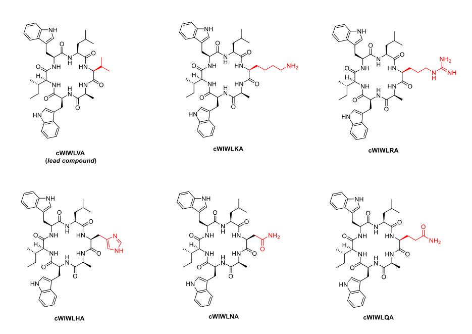
Figure 3 Nocardiotide-A (cWIWLVA) as lead compound and its analogues.
Radiopeptide on 99mTc requires that amino groups attach a chelating agent at the N-terminus of the peptide [17]. Nocardiotide-A consists of two aromatic amino acids of tryptophans and four hydrophobic amino acids, namely, alanine, valine, leucine, and isoleucine, where none of those amino acids have amine groups. Due to this, one of these amino acids was replaced by an amino group-possessing residue for binding to 99mTc. According to the previous studies by Muhajir et al. [8], the site between alanine at the C terminus and tryptophan at the N-terminal was selected for the cyclization of the linear precursor. This made the two residues irreplaceable and eventually made valine modification the most feasible way to make nocardiotide-A analogues. Therefore, nocardiotide-A analogues were designed by replacing valine with lysine (cWIWLKA), arginine (cWIWLRA), histidine (cWIWLHA), asparagine (cWIWLNA), and glutamine (cWIWLQA) individually, as shown in Figure 3.
Furthermore, to validate the binding pocket of cWIWLVA, cWIWLKA, cWIWLRA, cWIWLHA, cWIWLNA, and cWIWLQA, all peptides were docked onto SSTR2 as targets. Subsequently, a molecular dynamic simulation was performed for each peptide with the best value of free binding energy in SSTR2. The RMSD parameter analyzed the stability of each peptide complex, as shown in Figure 4. Based on the RMSD profile, the whole SSTR2 complex with nocardiotide-A analogues achieved stability starting at 80 ns of the simulation. Subsequently, no significant fluctuations were found indicating that the protein
remained stable during the 200-ns simulation. In the SSTR2 complex, cWIWLKA and cWIWLRA showed the best stability according to the lower RMSD value of each compound during the whole simulation.
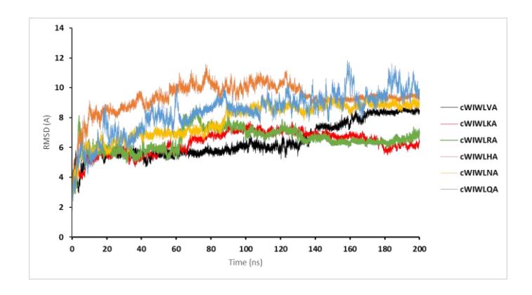
Figure 4 Plot of the RMSD value of nocardiotide-A and its analogues during the molecular dynamic simulation.
Also, the molecular dynamics trajectories of each peptide were visualized to observe their position and analyze their interaction. The snapshots at different times were taken for nocardiotide-A, as shown in Figure 5
Figure 5 The trajectory of nocardiotide-A against SSTR2.
The results of the binding pocket validation showed that the proposed binding site of nocardiotide-A analogues results in a docking area of 60 × 70 × 65 Å points centered on the x, y, and z coordinates of -15, 10, and 90 for cWIWLKA. For cWIWLHA, 52 × 67 × 70 Å points centered on the x, y, and z coordinates of -15, 10, and 90, 73 × 56 × 50 Å points centered on the x, y, and z coordinates of -15, 10, and 92 for cWIWLRA. Furthermore, 45 × 70 × 60 Å points centered on the x, y, and z coordinates of -15, 10, and 90 for cWIWLNA, while 74 × 50 × 50 Å points centered on the x, y, and z coordinates of -15, 21, and 90 for cWIWLQA.
The design of the radiopeptide based on 99mTc for molecular imaging consisted of a bifunctional chelator conjugated between peptide and 99mTc. The peptides are separated by a linker or spacer chain to avoid interference of the metal complex in the interaction with the target. 99mTc-HYNIC core has been widely used in radiopharmaceuticals. HYNIC has been used as bifunctional chelator, acting as mono or bidentate ligand, and requires co-ligands such as EDDA and tricine to complete the coordination of the Tc-HYNIC core [20].
Molecular docking calculations were performed to predict and understand the binding modes of the designed nocardiotide-A analogues and radiopeptide to the SSTR2 active site cavity using the technetium metal structural HYNIC model. The preferred conformations were selected by examining the cluster with the lowest mean binding energy. Table 1 shows the docking scores for the highestscoring cluster of poses. As modified peptides of nocardiotide-A, cWIWLKA, cWIWLRA, cWIWLHA, cWIWLNA and cWIWLQA had average docking scores when compared to cWIWLKA, the lead compound. All the nocardiotide-A analogues had negative free binding energies, indicating that they had good affinities to SSTR2.
In the complex system, hydrogen bonds and hydrophobic interactions play an essential role in protein-ligand interaction related to the binding affinity results. The biological activity of SSTR2 ligand with electronegative, less bulky, and hydrogen atom-donating/accepting substitutions is essential. D89, F92, Q102, A104, D122, N125, Q126, I.209, T212, G216, F265, W269, F272, Y.273, N276, K291, V.298, T.301, Y.302, N304, and S305 have been identified as crucial residues that form hydrogen bonds in the SSTR2 binding site [13]. Other critical SSTR2 residues that have been reported as vital to the bioactivity of ligands targeting SSTR2 are A104, K291, V298, and S305 [21]; D122 [22] and F294, D295, F296 and V297 [23].
Table 1 Docking nocardiotide-A and its analogues with SSTR2.
| No | Compound | ∆G (kcal/mol) | Hydrogen bond | Hydrophobic interaction |
|---|---|---|---|---|
| 1 | cWIWLVA | -10.41 | Q126, N276, S279 | F98, L99, M119, I177, F217, F275, F294 |
| 2 | cWIWLKA | -12.71 | Q102, D122, Y205, N276 | L99, V118, M119, I195, W197 |
| 99mTc-EDDA-HYNIC-cWIWLKA | - 8.61 | Q102 | L99, W188, I195, I209, F272, P286 | |
| 99mTc-tricine-HYNIC-cWIWLKA | - 3.12 | C193 | W108, V118, W188, Y205, A283, I284 | |
| 3 | cWIWLRA | -13.21 | V118, Y205, F275, N276 | I177, I209, F213, F217, F272, Y273, F294, V298 |
| 99mTc-EDDA-HYNIC-cWIWLRA | - 9.47 | D122, S279 | Y50, L96, L99, V103, V118, M119, V298 | |
| 99mTc-tricine-HYNIC-cWIWLRA | - 7.16 | N276, S279 | L99, V118, Y.205, V208, I209 | |
| 4 | cWIWLHA | -13.34 | Q102 | L99, R116, I117, V118, M119, A181, Y205, F208, I209, V280 |
| 99mTc-EDDA-HYNIC-cWIWLHA | - 9.05 | Q102, W197, S201 | V106, W108, A113, I195, M293, F275, | |
| 99mTc-tricine-HYNIC-cWIWLHA | - 3.34 | Q102 | V103, V106, I195, F275, M293 | |
| 5 | cWIWLNA | -12.56 | R184, C193, T.194, Y205, S279, F294 | L99, W108, M119, I195, F275, V298 |
| 99mTc-EDDA-HYNIC-cWIWLNA | -12.59 | E200, Y205, S281, M282 | W108, F208 | |
| 99mTc-tricine-HYNIC-cWIWLNA | - 8.96 | V106, N186 | W108, V118, M119, Y205, F208, I209, F275, V298, | |
| 6 | cWIWLQA | -6.56 | R184, C193, Y205, S279 | V106, W188, I195, S201 |
| 99mTc-EDDA-HYNIC-cWIWLQA | -6.17 | S279, I284 | V106, W188, I195, T194, Y205, A283, F294 | |
| 99mTc-tricine-HYNIC-cWIWLQA | -4.48 | Y37, Y38, S185, Q187, S279 | I195, F204, Y205 |
In these docking studies, the primary interaction between nocardiotide-A, its analogues, and SSTR2 had conventional hydrogen bonds. Nocardiotide-A and its analogues formed hydrogen bonds with Q102, D122, Q126, N276, and F294 residues. Also, nocardiotide-A and its analogues had hydrophobic interactions with I209, F272, Y273, F294, and V298 residues, which were identified as critical residues of the proposed binding site of SSTR2. According to this simulation, these ligand interactions that play a role in ligand binding may
influence the peptide's affinities to SSTR2. Figure 6 shows the ligand interaction of nocardiotide-A and its analogue in the active sites of SSTR2.
A molecular docking simulation of the 10 technetium metal structural HYNIC model of designed radiopeptides base showed that all radiopeptides had good binding affinity toward the active site of SSTR2. The in-silico study, according to Table 1, revealed that nocardiotide-A analogues conjugated to 99mTc-HYNIC-EDDA and 99mTc-HYNIC-tricine had hydrogen bonds with residues in the active site of SSTR2, including Q102, D122, and N276. All designed radiopeptides also had some hydrophobic interactions with residues in the proposed binding site, including those identified as critical residues of SSTR2, Y50, I209, F272, Y273, F294, and V298.
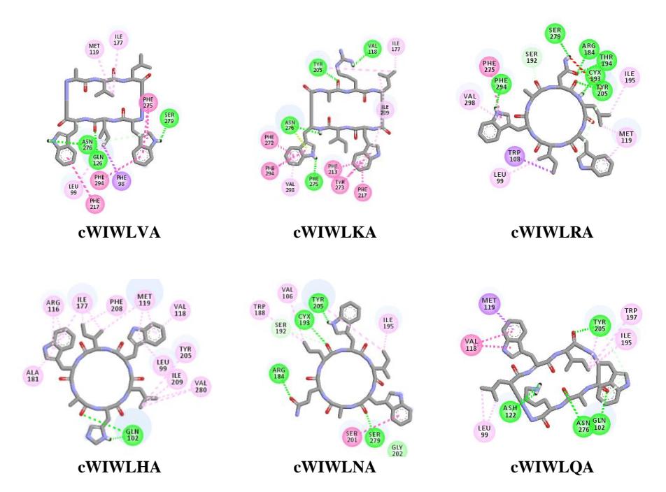
Figure 6 Interaction of nocardiotide-A and its analogue in SSTR2.
Although the interaction between the radiopeptides and a critical residue on the SSTR2 remained, the result showed that the binding affinity of the radiopeptides was much lower than that of the peptides. This means that the conjugation of peptide to 99mTc influences the binding affinity. Furthermore, the decrease in binding affinity is probably due to a slight change in the conformation position of the peptide because of the steric effect of the chelator/co-ligand and the partial charge of the molecule. A computational study also showed that the binding affinity of 99mTc/tricine/HYNIC-peptide decreased significantly compared to 99mTc/EDDA/HYNIC-peptide. The difference in docking score is probably due to the smaller size of 99mTc/EDDA/HYNIC compared to the 99mTc/tricine/HYNIC structure.
Figure S.3 shows the ligand interaction of the designed radiopeptides based on nocardiotide-A in the active sites of SSTR2.
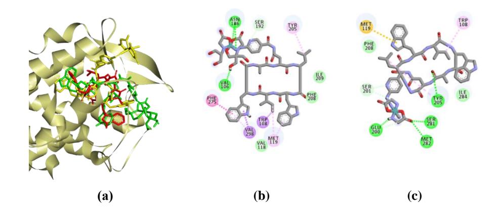
Figure 7 (a) Overlay of cWIWLNA (red) 99m Tc-EDDA-HYNIC-cWIWLNA (yellow) and 99m Tc-tricine-HYNIC-cWIWLNA (green) in the active sites of SSTR2 (b) Molecular interaction of 99m Tc - tricine -HYNIC-cWIWLNA, and (c) 99mTc- EDDA -HYNIC-cWIWLNA.
Among all the radiopeptides, 99mTc/EDDA/HYNIC-WIWLNA and 99mTc/tricine/HYNIC-WIWLNA had the best docking parameter with a free binding energy of -12.59 kcal/mol, and -8.96 kcal/mol, respectively. Figure 8 depicts the best pose of cWIWLNA, labeled by 99mTc, along with the significant amino acids contributing to the ligand-protein binding.
4 Conclusion
The computational study showed that all the modified nocardiotide-A and designed radiopeptides have binding affinity to SSTR2. According to the ligand interaction study of radiopeptide designed for nocardiotide-A analogues on 99mTc-core, the 99mTc/EDDA/HYNIC-peptide model was predicted to have a higher affinity for SSTR2 than that of the 99mTc/tricine/HYNIC-peptide model.
Acknowledgements
This study was supported partially by the Research Organization for Nuclear Energy, formerly the National Nuclear Energy Agency Scholarship Program Fiscal Year 2019-2021.