
1 Introduction
Cryptocarya, consisting of about 350 species, is a large genus of Lauraceae and is distributed in Asia, Australia, Africa, and South America [1]. It is locally known as medang in Indonesia and has been valued for a number of purposes, for example, as a source for building materials and furniture, pulp, and perfume [2,3]. Some of the plants have also been used in traditional medicines, for instance, for the treatment of muscle and joint pain, fever, headache, nausea, and infection caused by fungi [2−4]. The genus of this plant has been reported to produce a diverse range of secondary metabolites, including alkaloids, steroids, terpenoids, coumarins stilbenes, flavonoids, lignans, -pyrones, amides, and
Received January 6 th, 2023, Revised September 1 st, 2023, Accepted for publication June 12 th, 2024 Copyright © 2024 Published by ITB Institut for Research and Community Service, ISSN: 2337-5760, DOI: 10.5614/j.math.fund.sci.2024.55.3.5math
carboxylic acid derivatives. In a previous study [5], we have reported -pyrones and amides derivatives from Indonesian Cryptocarya that exhibited potential cytotoxic activity against murine leukemia P-388 cells. As a continuation of this study, herein we report the isolation and structure elucidation of compounds from Cryptocarya crassinervia leaves and their evaluation of inhibitory activity against receptor tyrosine kinases (RTKs).
2 Experiment
2.1 General
Gravity column chromatography (GCC) was carried out using two stationary phases, i.e., polyamide and Si gel 60 GF254. Meanwhile, centrifugal planar chromatography (CPC) was conducted using Si gel 60 PF254. Aluminum thinlayer chromatography (TLC) used Si gel coated with fluorescence indicator F254 (Merck Kieselgel 60 F254). Solvents (n-hexane, dichloromethane, ethyl acetate, and methanol) for extraction and purification were technical-grade and were distilled prior to use, except for chloroform, which was pro-analysis grade (Merck). 1H and 13C NMR spectra were measured with Agilent DD2 system operating at 500 (1H) and 125 (13C) MHz. High-resolution mass spectra (HRMS) were obtained with an ESI-TOF Waters LCT Premier XE. Optical rotations were measured with an Autopol IV Rudolph Research Analytical. Promega provided the ADP-Glo Kinase Assay and Kinase Selectivity Profiling System (KSPS) for receptor tyrosine kinases TK1 (EGFR, HER2, HER4, IGF1R, InsR, KDR, PDGFR, and PDGFR). The kinase enzyme assay was carried out using a Pipetmax-268 automatic liquid handler from Gilson, while the luminescent assay was measured with a GloMax Explorer.
2.2 Plant Materials
Leaves of C. crassinervia were collected from Bogor Botanical Garden, Bogor, West Java, Indonesia, in July 2017, and a voucher specimen (No. XX.B.12a) was deposited at the Herbarium Bogoriense.
2.3 Extraction and Isolation
The dried and powdered leaves of C. crassinervia (1.2 kg) were macerated with methanol at room temperature three times overnight (3 L each). After solvent evaporation, the extract (127 g) was separated from the chlorophyll by dissolving it in CH3OH−H2O (1:1). The free chlorophyll fraction (317.5 mL) was then redissolved in EtOAc to yield 5.5 g of EtOAc extract. The EtOAc extract was fractioned by using gravitational column chromatography (GCC; polyamide, 50 g) and eluted with CHCl3-CH3OH (9.5:0.5) to give 15 fractions (fr.A−fr.O).
Fraction B (1.4 g) was further subjected to centrifugal planar chromatography (CPC) with an eluent of CHCl3−acetone (7:3) and yielded compound 1 (18.9 mg). Fraction K (230 mg) was fractioned by GCC (eluent: CHCl3-CH3OH = 9:1) to give six fractions. Fraction K.4 was repeatedly separated using GCC eluted by CHCl3−CH3OH. Purification of the K4.2 fraction by CPC gave compound 2 (20 mg), while the K4-4 fraction afforded compound 3 (22 mg). Moreover, fraction M (380 mg) was further purified using GCC with CH3Cl−CH3OH as an eluent (9:1) to get fractions M.1−M.2 that were pure and then assigned as compound 4 (24.8 mg).
2.4 Tyrosine Kinase Assay
The tyrosine kinase assay was carried out in accordance with the established methodology previously described in [6], which is based on the work of Hennek [7], with some modifications. Specifically, the single-dose inhibition protocol was utilized to determine the percentage of enzyme activity. The test compound, prepared at a concentration of 5% in DMSO, was diluted with 62.5 μL of 4X kinase buffer and supplemented with 175 μL of nuclease-free water to achieve a concentration of 10 μM. Each kinase stock in the strip within the PCR tubes was diluted with 95 μL of 2.5X kinase reaction buffer, while each substrate/cofactor stock in the substrate strip was diluted with 20 μL of 80 μM ATP. These tubes were maintained at 0 °C passive thermal block until use. To initiate the assay, 2 μL of kinase working stocks and 2 μL of ATP/substrate mixture were dispensed into the wells of a 384-well plate along with 1 μL of the test compound. The reaction mixture was incubated for 1 hour at room temperature (22 to 25 °C).
Upon finishing the reaction, 5 μL of ADP-Glo reagent was added and incubated for another 40 minutes. Afterward, 10 μL of kinase detection reagent was added, and the incubation was continued for an additional 30 minutes to convert ADP to ATP, thus enabling the measurement of newly synthesized ATP through a luciferin reaction. Following the completion of the reaction, the luminescence was measured to the detected kinase activity. The negative control in absence of the compound represented uninhibited kinase activity (100% activity) and without the enzyme represented background luminescence (0% activity). The data were processed using the SMART protocol in the GloMax Explorer software. The reported percentage of kinase activity was calculated by subtracting the average luminescence of the no-enzyme control from all kinasecontaining reactions, with or without the compound, and then converting these net luminescence values to percentage activity based on the no-compound control reaction, representing 100% kinase activity. Erlotinib was used as positive control (1 μM).
3 Results and Discussion
The structure of the isolated compounds (1–4) was elucidated based on their spectroscopic data, including NMR, HRMS, and UV-Vis spectra, compared to the data in the literature.
Cryptocrassinervione (1) – Colorless gum: \([\alpha]_D^{20}\)-284 (c 0.05, MeOH); UV (MeOH) \(\lambda_{max}\) (log \(\epsilon\)): 204 (3.18) nm; IR (KBr) \(\nu_{max}\): 3451, 2928, 1713, 1641, 1387, 1258, 1103, 1038 cm<sup>-1</sup>; <sup>1</sup>H NMR (CDCl<sub>3</sub>) see Table 1. <sup>13</sup>C NMR (CDCl<sub>3</sub>) see Table 1. HRESITOF-MS m/z: \([M+Na]^+\) 381.1667 (calcd. for \(C_{21}H_{26}O_5Na\): 381.1672).
Afzelin (2) – Yellow solid: \([\alpha]_D^{26}\)-136 (c 0.05, MeOH); M.p. 175–176°C. UV (MeOH) \(\lambda_{\text{max}}\) (log \(\varepsilon\)): 219 (5.93), 265 (8.97), 296 (5.75), dan 339 (5.76) nm. <sup>1</sup>H NMR (500 MHz, acetone-\(d_6\)) \(\delta_{\text{H}}\): 12.68 (1H, s, 5-OH), 7.82 (2H, d, J = 8.7 Hz, H-2'/H-6'), 6.98 (2H, d, J = 8.7 Hz, H-3'/H-5'), 6.44 (1H, d, J = 1.8 Hz, H-8), 6.23 (1H, d, J = 1.9 Hz, H-6), 5.51 (1H, s, H-1"), 4.14 (1H, s, H-2"), 3.65 (1H, dd, J = 9.4 and 3.8 Hz, H-3"), 3.31 (1H, dd, J = 9.4 and 8.6 Hz, H-4"), 3.27 (1H, m, H-5"), 0.86 (3H, d, J = 5.8 Hz, H-6"); <sup>13</sup>C NMR (125 MHz, acetone-\(d_6\)) δ<sub>C</sub>: 179.1 (C-4), 164.8 (C-7), 163.0 (C-5), 160.7 (C-4'), 158.3 (C-2), 157.8 (C-8a), 135.5 (C-3), 131.5 (C-2'/C-6'), 122.3 (C-1'), 116.1 (C-3'/C-5'), 105.6 (C-4a), 102.5 (C-1"), 99.4 (C-6), 94.4 (C-8), 72.8 (C-4"), 72.0 (C-3"), 71.3 (C-2"), 71.2 (C-5"), 17.6 (C-6") ppm.
Quercitrin (3) – Yellow solid: \([\alpha]_D^{27,5}\)-76 (c 0.05, MeOH); <sup>1</sup>H NMR (500 MHz, methanol-\(d_4\)) \(\delta_{\rm H}\): 7.33 (1H, d, J = 1.9 Hz, C-2'), 7.29 (1H, dd, J = 8.3 and 1.9 Hz, H-6'), 6.90 (1H, d, J = 8.3 Hz, H-5'), 6.35 (1H, d, J = 1.6 Hz, H-8), 6.18 (1H, d, J = 1.8 Hz H-6), 5.34 (1H, d, J = 1.1 Hz, H-1"), 4.21 (1H, dd, J = 3.0 and 1.5 Hz, H-2"), 3.74 (1H, dd, J = 9.4 and 3.3 Hz, H-3"), 3.41 (1H, m, H-5"), 3.33 (1H, d, J = 9.5 Hz, H-4"), 0.94 (3H, d, J = 6.1 Hz, H-6") ppm; <sup>13</sup>C NMR (125 MHz, methanol-\(d_4\)) \(\delta_{\rm C}\): 179.6 (C-4), 165.9 (C-7), 163.2 (C-5), 159.3 (C-2), 158.5 (C-8a),149.8 (C-4'), 146.4 (C-3'), 136.2 (C-3), 123.0 (C-1'), 122.9 (C-6'), 116.9 (C-2'), 116.4 (C-5'), 105.9 (C-4a), 103.5 (C-1"), 99.8 (C-6), 94.7 (C-8), 73.3 (C-4"), 72.1 (C-5"), 72.0 (C-3"), 71.9 (C-2"), 17.7 (C-6") ppm.
Kaempferol-3-O-rhamnosyl-2-O-apiofuranoside (4) – Yellowish powder: \([\alpha]_D^{25}\)-56 (c 0.05, MeOH); <sup>1</sup>H NMR (acetone-\(d_6\)) see Table 2; <sup>13</sup>C NMR (acetone-\(d_6\)) see Table 2. HRESITOF-MS m/z: \([M-H]^-\) 563.1409 (calcd. for \(C_{26}H_{27}O_{14}\): 563.1401).
Among the four compounds obtained from the leaf extract of C. crassinervia (Figure 1), two were new, i.e., cryptocrassinervione (1), a new \(\alpha\)-pyrone, and kaempferol-3-O-rhamnosyl-2-O-apiofuranoside (4), a new flavonoid glycoside. Besides that, two known flavonoid glycosides, namely afzelin (2) and quercitrin (3), were also isolated.
OH OH \[A_1\] \(A_2\) \(A_3\) \(A_4\) \(A_4\) \(A_4\) \(A_4\) \(A_4\) \(A_4\) \(A_4\) \(A_4\) \(A_4\) \(A_4\) \(A_4\) \(A_4\) \(A_4\) \(A_4\) \(A_4\) \(A_4\) \(A_4\) \(A_4\) \(A_4\) \(A_4\) \(A_4\) \(A_4\) \(A_4\) \(A_4\) \(A_4\) \(A_4\) \(A_4\) \(A_4\) \(A_4\) \(A_4\) \(A_4\) \(A_4\) \(A_4\) \(A_4\) \(A_4\) \(A_4\) \(A_4\) \(A_4\) \(A_4\) \(A_4\) \(A_4\) \(A_4\) \(A_4\) \(A_4\) \(A_4\) \(A_4\) \(A_4\) \(A_4\) \(A_4\) \(A_4\) \(A_4\) \(A_4\) \(A_4\) \(A_4\) \(A_4\) \(A_4\) \(A_4\) \(A_4\) \(A_4\) \(A_4\) \(A_4\) \(A_4\) \(A_4\) \(A_4\) \(A_4\) \(A_4\) \(A_4\) \(A_4\) \(A_4\) \(A_4\) \(A_4\) \(A_4\) \(A_4\) \(A_4\) \(A_4\) \(A_4\) \(A_4\) \(A_4\) \(A_4\) \(A_4\) \(A_4\) \(A_4\) \(A_4\) \(A_4\) \(A_4\) \(A_4\) \(A_4\) \(A_4\) \(A_4\) \(A_4\) \(A_4\) \(A_4\) \(A_4\) \(A_4\) \(A_4\) \(A_4\) \(A_4\) \(A_4\) \(A_4\) \(A_4\) \(A_4\) \(A_4\) \(A_4\) \(A_4\) \(A_4\) \(A_4\) \(A_4\) \(A_4\) \(A_4\) \(A_4\) \(A_4\) \(A_4\) \(A_4\) \(A_4\) \(A_4\) \(A_4\) \(A_4\) \(A_4\) \(A_4\) \(A_4\) \(A_4\) \(A_4\) \(A_4\) \(A_4\) \(A_4\) \(A_4\) \(A_4\) \(A_4\) \(A_4\) \(A_4\) \(A_4\) \(A_4\) \(A_4\) \(A_4\) \(A_4\) \(A_4\) \(A_4\) \(A_4\) \(A_4\) \(A_4\) \(A_4\) \(A_4\) \(A_4\) \(A_4\) \(A_4\) \(A_4\) \(A_4\) \(A_4\) \(A_4\) \(A_4\) \(A_4\) \(A_4\) \(A_4\) \(A_4\) \(A_4\) \(A_4\) \(A_4\) \(A_4\) \(A_4\) \(A_4\) \(A_4\) \(A_4\) \(A_4\) \(A_4\) \(A_4\) \(A_4\) \(A_4\) \(A_4\) \(A_4\) \(A_4\) \(A_4\) \(A_4\) \(A_4\) \(A_4\) \(A_4\) \(A_4\) \(A_4\) \(A_4\) \(A_4\) \(A_4\) \(A_4\) \(A_4\) \(A_4\) \(A_4\) \(A_4\) \(A_4\) \(A_4\) \(A_4\) \(A_4\) \(A_4\) \(A_4\) \(A_4\) \(A_4\) \(A_4\) \(A_4\) \(A_4\) \(A_4\) \(A_4\) \(A_4\) \(A_4\) \(A_4\) \(A_4\) \(A_4\) \(A_4\) \(A_4\) \(A_4\) \(A_4\) \(A_4\) \(A_4\) \(A_4\) \(A_4\) \(A_4\) \(A_4\) \(A_4\) \(A_4\) \(A_4\) \(A_4\) \(A_4\) \(A_4\) \(A_4\) \(A_4\) \(A_4\) \(A_4\) \(A_4\) \(A_4\) \(A_4\) \(A_4\) \(A_4\) \(A_4\) \(A_4\) \(A_4\) \(A_4\) \(A_4\) \(A_4\) \(A_4\) \(A_4\) \(A_4\) \(A_4\) \(A_4\) \(A_4\) \(A_4\) \(A_4\) \(A_4\) \(A_4\) \(A_4\) \(A_4\) \(A_4\) \(A_4\) \(A_4\) \(A_4\) \(A_4\) \(A_4\) \(A_4\) \(A_4\) \(A_4\) \(A_4\) \(A_4\) \(A_4\) \(A_4\) \(A_4\) \(A_4\) \(A_4\) \(A_4\) \(A_4\) \(A_4\) \(A_4\) \(A_4\) \(A_4\) \(A_4\) \(A_4\) \(A_4\) \(A_4\) \(A_4\) \(A_4\) \(A_4\) \(A_4\) \(A_4\) \(A_4\) \(A_4\) \(A_4\) \(A_4\) \(A_4\) \(A_4\) \(A_4\) \(A_4\) \(A_4\) \(A_4\) \(A_4\)
Figure 1 Structures of compounds isolated from C. crassinervia.
Compound 1 was isolated as a yellowish gum, \([\alpha]_D^{20}\)-284° (MeOH), and has the molecular formula of C21H26O5 by high resolution electrospray ionization-time of flight mass spectrometry (HR-ESI-TOF-MS) (found m/z [M+Na]<sup>+</sup> 381.1667, calcd. 381.1672). The UV spectrum of 1 showed the maximum absorption at \(\lambda_{max}\) 204 nm, which is the typical absorption of a simple benzenoid chromophore. Meanwhile, its IR spectrum showed absorptions at \(v_{\text{max}}\) 3451 (OH) and 1713 (conjugated ester C=O) cm<sup>-1</sup>. These IR data indicated that compound 1 is an \(\alpha\)-pyrone derivative [8]. In the NMR spectra (Table 1), the \(\alpha\)-pyrone structure is shown by the presence of signals for a conjugated carbonyl lactone C-2 (δ<sub>C</sub> 164.4), a conjugated cis-disubstituted alkene (\(\delta_{\rm H}\) 6.86 (H-3), 6.02 (H-4), each J = 9.7 Hz; \(\delta_{\rm C}\) 121.4 (C-3), 145.1 (C-4)), a methylene at C-5 (\(\delta_H\) 2.37, \(\delta_C\) 28.6), and an oxymethine at C-6 (\(\delta_H\) 4.60, \(\delta_C\) 75.2) groups, which was confirmed by the HMBC correlations (Fig. 2). The NMR spectra also showed the signals at \(\delta_H\) 7.28 (H-3"/H-5"), 7.19 (H-4"), 7.17 (H-2"/H-6"), 2.86, (H-10'), 2.76 (H-9'), and signal at \(\delta_C\) 207.8 (C-8') that indicated the presence of a dihydrocinnamoyl group. Meanwhile, the NMR signal at \(\delta_H\) 4.23 (H-2'), 4.16 (H-6'), 4.05 (H-4'), 2.80 & 2.51 (H-7'), 2.29 & 1.71 (H-1'), 1.80 & 1.64 (H-3'), and 1.94 & 1.29 (H-5') exhibited a 2,6-dimethylenepyran-4-ol moiety.
The selected important HMBC showed the correlation between methylene at C-7' of the 2,6-dimethylenepyran-4-ol moiety (\(\delta_{\rm H}\) 2.80 & 2.51 ppm) and the carbonyl of a dihydrocinnamoyl at \(\delta_{\rm C}\) 207.8 ppm. Besides that, there was also an HMBC correlation between methylene at C-1' (\(\delta_{\rm H}\) 1,71 and 2,29 ppm) and C-6 of a pyrone ring (\(\delta_C\) 75.2 ppm). From this analysis, the structure of 1 can be formulated as a cinnamoyl unit and an \(\alpha\)-pyrone unit flanking the 2,6-dimethylenepyran-4-ol moiety.
Long-range \(^1H^{-13}C\) correlations (Figure 2) between the methylene proton signals at \(\delta_H\) 2.80 & 2.51 and 2.29 & 1.71 of the 2,6-dimethylenepyran-4-ol moiety with the carbon signals at \(\delta_C\) 207.8 and 75.2, respectively, confirmed the basic structure of 1. Other HMBC correlations that support structure 1 are shown in Fig. 2. Compound 1 is, therefore, an 8,9-dihydroderivative of cryptoconcatone, which was isolated from C. concinna [9]. The CD spectrum revealed positive Cotton effects at \(\lambda\) (\(\Delta\epsilon\)) 205 (+32.19), 236 (+6.69), and 254 (+10.09) nm, consistent with the R configuration, indicating absolute stereochemistry at C-6 [8]. The presence of large coupling constants in the H-2' and H-6' signals (9.5 and 9.1 Hz, respectively) supported by NOE interaction (Figure 3) indicate that these hydrogens are in the axial configuration, and therefore the oxygen functionalities in the pyran-4-ol ring are equatorial. Therefore, compound 1 is trivially named as cyrptocrassinervione (Figure 1).
Compound 4 was isolated as a pale-yellow solid, \([\alpha]_D^{20}\)-56° (MeOH). Based on the HRESITOF-MS spectrum (negative mode), it revealed a quasimolecular ion [M-H]<sup>-</sup> at m/z 563.1409, consistent with the molecular formula \(C_{26}H_{28}O_{14}\) (calcd. [M-H]<sup>-</sup> at m/z 563.1401). The <sup>1</sup>H NMR data of compound 4 (Table 2) were similar to those of compound 2, namely the signals for the kaempferol structure, including signals of two protons of tetrasubstituted benzene at \(\delta_H\) 6.46 (H-6) & 6.26 (H-8), each 1H and d, J = 2.0 Hz. In addition, there were also signals at \(\delta_H\) 7.84 (H-2'/H-6') & 7.02 (H-3'/H-5'), each 2H and d, J = 8.7, indicating the presence of disubstituted benzene. Moreover, there were also signals for the α-L-rhamnopyranosyl groups at \(\delta_H\) 5.55 (H-1"), 4.23 (H-2"), 3.80 (H-3"), 3.44 (H-5"), 3.34 (H-4"), and 0.94 (H-6"). Compound 4 also had additional NMR signals that appeared at \(\delta_H\) 5.19 (H-1""), 3.97 (H-2""), 3.82 & 3.69 (H-4""), and 3.56 (H-5""), as well as signals at \(\delta_C\) 111.5 (C-1""), 80.0 (C-3""), 77.3 (C-2""), 74.7 (C-4""), and 65.6 (C-5""), that determined the presence of β-D-apiofuranosyl [13].
The HMBC spectrum showed a correlation between the anomeric proton signal of the \(\beta\)-D-apiofuranosyl group (\(\delta_H\) 5.19, d, J=1.9 Hz) and the carbon signal at C-2 (\(\delta_C\) 77.9) that indicated that the \(\beta\)-D-apiofuranosyl group attached at C-2 of \(\alpha\)-L-rhamnopyranosyl group. Thus, 4 was determined as kaempferol-3-O-(2-O-\(\beta\)-D-apiofuranosyl)-\(\alpha\)-L-rhamnopyranoside. Further support for structure 4 was obtained by comparison of the NMR data of 4 with those reported for quercetin-3-O-(2-O-\(\beta\)-D-apiofuranosyl)-\(\alpha\)-L-rhamno-pyranoside [10].
| C No. | 1 | 4 | |||
|---|---|---|---|---|---|
| H (m, J in Hz) | C | C No. | H (m, J in Hz) | C | |
| 2 | - | 164.4 | 2 | - | 158.3 |
| 3 | 6.02 (ddd, 9.7,1.9, 1.0) | 121.4 | 3 | - | 135.7 |
| 4 | 6.86 (ddd, 9.7, 5.6, 2.9) | 145.1 | 4 | - | 179.1 |
| 5 | 2.37 (m) | 28.6 | 4a | - | 105.6 |
| 6 | 4.60 (ddt, 10.5, 7.5, 5.2) | 75.2 | 5 | - | 163.1 |
| 1' | 1.71 (ddd, 14.4, 7.5, 4.7) | 36.7 | 6 | 6.26 (d, 2.0) | 99.4 |
| 2.29 (ddd, 14.4, 9.9, 5.2) | - | 7 | - | 164.8 | |
| 2' | 4.23 (dq, 9.5, 4.7) | 67.0 | 8 | 6.46 (d, 2.0) | 94.4 |
| 3' | 1.64 (ddd, 13.1, 9.5, 5.2) | 37.9 | 8a | - | 157.9 |
| 1.80 (dtd, 13.1, 4.7, 1.6) | - | 1' | - | 122.3 | |
| 4' | 4.05 (br q, 4.7) | 64.0 | 2'/6' | 7.84 (d, 8.7) | 131.5 |
| 5' | 1.29 (dt, 12.8, 9.2) | 39.4 | 3'/5' | 7.02 (d, 8.7) | 116.3 |
| 1.94 (br dt, 12.8, 3.5) | - | 4' | - | 160.8 | |
| 6' | 4.16 (tdd, 9.1, 4.6, 3.5) | 65.8 | 1'' | 5.55 (d, 1.1) | 101.8 |
| 7' | 2.80 (dd, 16.1, 9.1) | 48.3 | 2'' | 4,23 (dd, 3.0, 1,6) | 77.9 |
| 2.51 (dd, 16.1, 4.6) | - | 3'' | 3.80 (dd, 3.1, 9.3) | 71.7 | |
| 8' | - | 207.8 | 4'' | 3.34 (t, 9.5) | 73.3 |
| 9' | 2.76 (t, 7.3) | 45.2 | 5'' | 3.44 (m) | 71.3 |
| 10' | 2.86 (t, 7.3) | 29.5 | 6'' | 0.94 (d, 6.1) | 17.8 |
| 1'' | - | 140.9 | 1''' | 5.19 (d, 1.9) | 111.5 |
| 2''/6'' | 7.17 (br d, 7.4) | 128.3 | 2''' | 3.97 (d, 1.6) | 77.3 |
| 3''/5'' | 7.28 (br d, 7.4) | 128.5 | 3''' | - | 80.0 |
| 4'' | 7.19 (br t, 7.4) | 126.2 | 4''' | 3.82 (d, 9.6) | 74.7 |
| 126.2 | 3.69 (d, 9.6) | ||||
| 126.2 | 5''' | 3.56 (s) | 65.6 | ||
| 126.2 | 5-OH | 12.73 (brs) | - | ||
Table 1 NMR data of compounds 1 (CDCl3) and 4 (acetone-d6).
The 13C NMR spectrum of 2, identified as afzelin (kaempferol-3-O--Lrhamnoside), showed the presence of 19 signals representing 21 carbon atoms, six of which were signals for C-sp3 . The remaining 13 carbon signals were for C-sp2 , including two characteristic carbon signals for a flavonol derivative at δc 179.1 and 135.5. The 13C NMR spectrum indicated that molecule 2 was likewise a glycosylated flavonol derivative. The 1H NMR spectrum also revealed signals corresponding to the kaempferol unit, which was the same as in compound 4. The signals included two ortho-coupled aromatic doublet signals (δH 7.82 and 6.98) on ring B, two meta-coupled aromatic doublet signals (δH 6.23 and 6.44) on ring A and a chelated OH group proton signal (δH12.68). The existence of a methyl proton doublet signal at δH 0.86 showed the glycoside group in 2 originates from an α-L-rhamnosyl group, similar to compound 4. The NMR data for 2 was compared to the data for the same compound in the literature and showed good agreement [11].
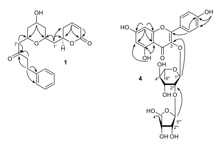
Figure 2 Selected important HMBC (1H 13C) correlations in 1 and 4.
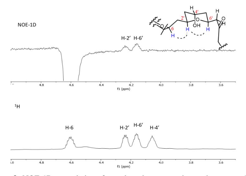
Figure 3 NOE-1D correlation for selected protons inset the pyran-4-olconformation of 1.
Whereas the 1H NMR spectrum of 3, namely quercitrin (quercetin-3-O--Lrhamnoside), was identical to compound 4, with a pair of meta-coupled aromatic
proton signals on ring A at δH 6.35 and 6.18 and the characteristic carbon signals for flavonol derivatives at δC 179.6 and 136.2. Compound 3 also exhibited signals for the α-L-rhamnosyl group at δH 5.34, 4.21, 3.74, 3.33, 3.41, and 0.94. The difference was that the pair of ortho-coupled aromatic proton signals on ring B in compound 4 were replaced by three aromatic signals at δH 7.33, 6.90, and 7.29 in compound 3, with each having the multiplicity of d (J = 1.9 Hz), d (J = 8.3 Hz) and dd (J = 1.9 and 8.3 Hz), respectively, suggesting that the flavonol unit in compound 3 is quercetin. All the spectroscopic data were in accordance with the data in the literature for the same compound [12].
Furthermore, compounds 1−4 (at a concentration of 10 M) were evaluated against eight receptor tyrosine kinases (RTKs), including EGFR, HER2, HER4 (epidermal growth factor receptor), IGF1R, InsR (insulin receptor), KDR (kinase insert domain receptor), and PDGFR-α and - (platelet-derived growth factor receptor) [6]. The results showed that all compounds are selectively active against EGFR (moderate, inhibition percentage of 41−55%) and HER2 (weak, inhibition percentage of 15−20%) (Table 3). EGFR and HER2 are members of the epidermal growth factor receptor (EGFR) family involved in cell proliferation, anti-apoptosis, and tumor cell motility [13]. EGFR overexpression has been observed in non-small cell lung cancer (NSCLC), colorectal cancer (CRC), and squamous cell carcinoma of the head and neck (SCCHN), while HER2 mutations has been detected in patients with breast, gastroesophageal, pancreatic, and bladder cancer [14].
Table 2 Percent inhibition of 1−4 (at a concentration of 10 M) against eight receptor tyrosine kinasesa.
| Compds. | EGFR | HER2 | HER4 | IGF1R | InsR | KDR | PDGFRα | PDGFRβ |
|---|---|---|---|---|---|---|---|---|
| Erlotinibb | 100 | 94 | 77 | 0 | 2 | 96 | 87 | 87 |
| 1 | 55 | 17 | 0 | 0 | 0 | 0 | 3 | 0 |
| 2 | 49 | 20 | 0 | 4 | 0 | 0 | 2 | 2 |
| 3 | 41 | 18 | 0 | 4 | 0 | 0 | 2 | 0 |
| 4 | 44 | 15 | 0 | 0 | 0 | 19 | 11 | 0 |
a strong: >80%, moderate: 40−80%, weak or not active: <40% ; b positive control (1.0 M)
According to Safe et al. (2021) [15], flavonoids can bind to various proteins, including EGFR, and modify their function. Genistein (an isoflavone) is one of the flavonoids that has so far been recognized as an inhibitor of receptor tyrosine kinase. Therefore, our result, which showed that two flavonoids, i.e., compounds 2−4, have a moderate effect on EGFR, also supported that fact. From these results, it can be stated that the compounds isolated from leaf extract of C. crassinervia, have the potential to be lead compounds for anticancer agents through the inhibition of EGFR and HER2.
In conclusion, one new -pyrone (cryptocrassinervione (1)) and one new flavonol glycoside (quercetin-3-O-(2-O-β-ᴅ-apiofuranosyl)--L-rhamnopyranoside (4)), along with two known flavonol glycosides (afzelin (2) and quercitrin (3)), were isolated from leaf extract of C. crassinervia. All compounds were moderately active against EGFR but weakly active against HER2.
Acknowledgements
We dedicate this paper to the memory of our colleague, Prof. Yana Maolana Syah, who passed away while this paper was being revised. Prof. Syah contributed significantly to the preparation of the original manuscript. The scientific community also feels lost and deeply saddened by his demise.
We are grateful to BUDI DN 2016 LPDP-Ristekdikti for the scholarship and acknowledge the PDUPT (Penelitian Dasar Unggulan Perguruan Tinggi) 2017 from the Ministry of Research, Technology and Higher Education of the Republic of Indonesia for part of the financial support for this research.