
1. Introduction
Type 2 diabetes mellitus (T2DM) is often referred to as a "silent killer" because many individuals are unaware that they have the condition, increasing the risk of serious health complications. Globally, diabetes affects approximately 422 million people and contributes to about 1.5 million deaths each year [1]. In Indonesia, up to 90% of the population consume sweet food and beverages [2], even though the daily sugar intake recommended by Ministry of Health Regulation No. 30/2013 is limited to 50 g per person. This leads to an increase of diabetes cases in Indonesia by 42% in 2045 to 28.57 million people, compared to 2021, where active cases were numbered at 19.47 million diabetics with 57.42 deaths per 100 thousand Indonesians [3].
T2DM is a chronic metabolic disease with the main complication being disturbances in the carbohydrate metabolism system due to an increase in blood sugar. This may cause insulin resistance; the inability of the human body to respond well to insulin [4]. Insulin is a hormone produced in the human body that is crucial in cellular glucose uptake as a source of energy. If human cells become resistant to insulin, glucose will accumulate in blood and cause hyperglycaemia; which is a characteristic of T2DM [5]. Diabetes could be managed by inhibiting the enzymatic activity of α-amylase. α-Amylase degrades and hydrolyses polysaccharides (i.e. starch) into shorter chains of dextrin by breaking α-1,4 glycosidic linkages in polysaccharides [6]. Complex carbohydrates converted into simple sugars are absorbed in the small intestine and enter the bloodstream as energy. By inhibiting α-amylase, the production of glucose can be reduced, thus controlling blood sugar levels [7].
Quercetin (C15H10O7 ) is a bioactive flavonoid secondary metabolite predominantly found in plant matters; mainly vegetables and fruits [8]. Prior research shows the various health benefits of quercetin as an anti-inflammatory, antioxidant, antiviral, anti-cancer, and anti-diabetic agent [9]. By inhibiting α-amylase, quercetin could be utilized as a 308 DOI: 10.5614/3bio.2025.7.1.3
therapeutic drug to treat T2DM. Quercetin inhibits α-amylase by forming hydrogen bonds with the active sites of the enzyme; this mechanism of action is similar with common anti-diabetic drugs [10]. Pure quercetin has an IC50 value of 57.37 μg/mL in inhibiting α-amylase [7]. Previous research has proved that administering quercetin with daily dose of 20.9 ± 2.32 mg could reduce diabetes mellitus prevalence in the local population in China [11].
At present, diabetes is commonly treated by insulin therapy, oral drugs, and herbal medicines. Insulin and oral anti-diabetic drugs have been the most predominantly used agents in managing T2DM [12, 13]. However, the administering of insulin and medications often have adverse side effects on the human body. Long-term usage of insulin can lead to hypoglycaemia; a condition of low blood sugar levels. Hypoglycaemia has been the cause of death on 3% of T2DM patients that rely on insulin [13]. One of the most widely used anti-diabetic drugs is acarbose. Acarbose works by competitively inhibiting α-amylase, thus preventing the enzyme from hydrolysing starch into glucose. Yet, acarbose could cause side effects in the human gastrointestinal system (e.g. diarrhoea, flatulence, bloating) [14]. An alternative in treating diabetes is by administering herbal medicines with little to no side effects upon consumption [13]. Quercetin, that can be found in numerous plant matters, has a similar α-amylase inhibitory mechanism compared to acarbose and may potentially be used in T2DM treatment.
Shallot (Allium cepa L.) is a common agricultural commodity with high secondary metabolite contents (triterpenoids, phenolic acids, flavonoids, thiosulphates, anthocyanins) [15]. Shallot also contains phenols such as quercetin, isoquercetin, kaempferol, and isorhamnetin [16]. On average, a shallot contains 2.13 mg/mL of gallic acid, 35.91 mg/mL of quercetin, 0.66 mg/mL of kaempferol, and 21.71 mg/mL of tannic acid [17]. Flavonols, including quercetin, are the main substance of a shallot skin's abaxial. Quercetin was first isolated and identified as a flavonoid pigment in Allium cepa L. samples back in 1965 [18]. As a major flavonol compound found in Allium cepa L. cultivars, quercetin is predominantly present as quercetin 3,4′-diglucoside and quercetin 4′-monoglucoside [19]. This results in a higher level of flavonoids on the outer layers of the shallot compared to the inner layers of the bulb, with the highest flavonoid concentration located in the skin [20, 21]. A higher concentration of quercetin is needed as a protective measure; to defend itself from hydroxyl radicals, plants would produce antioxidative compounds on its peripheries [22]. Shallot wastes contain more bioactive compounds than the edible bulb itself [23]. Quercetin-rich skin accounts for more than 80% of the total flavonoid content in shallots. Shallot skin contains 6.5% (w/w) of quercetin, while the bulb itself only contains 0.01% (w/w) [24].
In Indonesia, shallot is widely used as a cooking ingredient. Farmers have cultivated onions for decades to fulfil an everincreasing demand by the market. In 2023, market demands for shallots in Indonesia reached a staggering level of 1,985,233 tonnes [25]. The increase in shallot production is followed by an increasing amount of shallot skin wastes. Untreated shallot skin wastes will produce foul odour caused by the formation of greenhouse gasses (e.g. methane, carbon dioxide) during its decomposition [26]. These quercetin-rich skin wastes can be used as raw materials for quercetin extraction. Using shallot skin waste to extract quercetin could solve the underutilization problem and provide a therapeutic alternative for T2DM. This research aims to determine the quercetin yield of Allium cepa L. skin waste, evaluate the α-amylase inhibitory properties of the extracted quercetin, and delve into the molecular mechanism of α-amylase inhibition by quercetin.
2. Methodology
2.1. Sample Collection and Preparation
Dried shallot skin was collected from the production wastes of fried shallot condiment. Then, the wastes were frozen at -20°C for three days in an air-tight container. The frozen wastes were then grounded into fine powder to be extracted [27].
2.2 Quercetin Extraction.
Finely ground shallot skin wastes were macerated in 70% ethanol for 24 hours. The maceration container was agitated for the first 3-6 hours to dissolve bioactive compounds contained inside the skin. Then, the blend was filtrated using Buchner funnel with Whatman No. 1 125 mm filter paper; this process was repeated three times. After filtration, the bioactive compound-rich solvent is evaporated inside a vacuum rotary evaporator [28]. The evaporation process was conducted in two stages; ethanol evaporation was done at a temperature of 65°C, followed by water evaporation at 75°C.
2.3 Preparation of Quercetin Standard Curve by Spectrophotometry
A quercetin standard curve was made using methods adopted from previous research [29] with slight modifications. First, 20 mg of standard quercetin (acquired from Sigma-Aldrich) was dissolved in 20 mL of pro analysis-grade methanol to produce 1000 µg/mL quercetin solution. The solution was further diluted to produce 100 µg/mL stock solution. Using the stock solution, standard sample solutions were prepared with concentrations of 20, 40, 60, 80, and 100 µg/mL. Then, 1 mL of standard sample from each concentration was added with 1 mL of 10% AlCl3 and 1 mL of 5% CH3COOH before being incubated for 30 minutes. The absorbance of the assay was then measured using a UV-Vis spectrophotometer with a wavelength of 433 nm in four replications per concentration.
2.4 Determination of Quercetin Levels in Extract by Spectrophotometry
The method to determine quercetin content in the shallot skin extract was adopted from previous research [29] with slight modifications. First, 20 mg of shallot skin extract was dissolved in 20 mL of pro analysis-grade methanol to produce 1000 µg/mL of extract solution. The solution was further diluted to acquire 100 µg/mL stock solution. Then, 1 mL of the stock solution was added with 1 mL of 10% AlCl3 and 1 mL of 5% CH3COOH before being incubated for 30 minutes. The absorbance of the assay was then measured using a UV-Vis spectrophotometer with a wavelength of 433 nm in sixteen replications.
2.5 Preparation of Quercetin Standard Curve by High-Performance Liquid Chromatography
Fifty mg of standard quercetin (acquired from Sigma-Aldrich) was dissolved in 10 mL of HPLC-grade methanol to produce 5000 µg/mL quercetin solution. It was then further diluted to produce 1000 µg/mL of quercetin stock solution. From the stock solution, standard sample solutions were prepared with concentrations of 10, 20, 40, 60, 80, and 100 µg/ mL. The analysis was done using Shimadzu high-performance liquid chromatograph with Nucleosil, Phenomenex, 5 μm C18 cartridge and LC Column 250 x 4.6 mm. The HPLC assay was carried out at a temperature of 25°C, a flow rate of 1 mL/min, a mobile phase of HPLC-grade methanol and 0.1% H3PO4 (55:45 v/v), and a detection wavelength of 370 nm in four replications per concentration. The volume used in each injection was 20 μL.
2.6 Determination of Quercetin Levels in Extract by High-Performance Liquid Chromatography
Ten mg of shallot skin extract was dissolved in 10 mL of HPLC-grade methanol to produce 1000 µg/mL of extract solution. The solution was further diluted to create three 50 µg/ mL sample solutions. The analysis was done using Shimadzu high-performance liquid chromatograph with Nucleosil, Phenomenex, 5 μm C18 cartridge and LC Column 250 x 4.6 mm. The HPLC assay was carried out at a temperature of 25°C, a flow rate of 1 mL/min, a mobile phase of HPLCgrade methanol and 0.1% H3PO4 (55:45 v/v), and a detection wavelength of 370 nm in sixteen replications. The volume used in each injection was 20 μL.
2.7 Determination of the α-Amylase Inhibitory Properties of Quercetin
The method for determining the α-amylase inhibitory was adopted from previous research [30] with slight modifications. First, 0.5% (w/v) potato starch solution was boiled for 15 minutes. Then, an enzyme solution was prepared by dissolving 10 mg of porcine pancreas α-amylase in 1 L of 20 mM phosphate buffer (pH 6.9). Sample solutions were made by dissolving shallot skin extract and acarbose in dimethyl sulfoxide (DMSO) solvent, with concentrations of 200, 400, 600, 800, and 1000 µg/mL. Ten mL of each concentration were made. Absorbance was measured for sample, blank, and control solutions. To make samples, 1 mL of each extract/acarbose concentration is mixed with 1 mL of enzyme solution and incubated at a temperature of 25°C for 30 minutes. Then, 1 mL of the mixture was taken and added into a reaction tube containing 1 mL of potato starch solution before being incubated at 25°C for 3 minutes. To stop the enzymatic reactions, 1 mL of DNS was added and the mixture is heated at 85°C for 15 minutes. After the reaction tube has cooled, 9 mL of distilled water was added into the tube. The mixture was then homogenized and had its absorbance measured using UV-Vis spectrophotometer at a wavelength of 540 nm. The absorbance measurement of blanks used the same method except for the order of addition; DNS was added before potato starch to prevent any enzymatic reactions from happening. Control absorbance was acquired by substituting extract or acarbose samples with 1 mL of DMSO. Each variation of solutions used were measured in four replications per concentration.
2.7.1 α-Amylase Inhibitory capability of Quercetin
Half maximal inhibitory concentration (IC50) is the concentration of a substance needed to inhibit a biochemical reaction by 50%. The IC50 value of a compound could be deduced from the linear regression between the concentration of the compound and its inhibition percentage. The inhibition percentage of porcine pancreas α-amylase is needed to calculate the amount of α-amylase that had been inhibited by quercetin, thus ascertaining its inhibitory capability. In this research, the measured inhibition of α-amylase by quercetin extract and acarbose is the product of prolonged incubation (i.e. 30 minutes). The end-point α-amylase inhibitory percentages and end-point IC50 values could be calculated by processing absorbance data using these formulas [28].
\[%Inhibition = \frac{Abs_{control} - (Abs_{sample} - Abs_{blank})}{Abs_{control}} \times 100\%\]
Abscontrol : control absorbance Abssample : sample absorbance Absblank : blank absorbance
\[IC_{50} = \frac{0.5 - b}{a}\]
For a general regression equation of y = ax + b.
310 DOI: 10.5614/3bio.2025.7.1.3
3. Results and Discussion
3.1 Extraction Result
The shallot samples had been identified as Allium cepa L. var. Sumenep by Herbarium Bandungense as per plant determination letter 5063/IT1.C11.2/TA.00/2024. Shallot skin wastes were procured from two home-based fried shallot businesses in Bandung, West Java. Wastes weighing 2.5 kg were acquired from Sedap Rasa production house, while 2.4 kg of wastes were acquired from Sari Asih production house. Waste samples that were used were dried shallot skin wastes with little to no contaminants. The wastes were then sealed in airtight plastic bags using a vacuum sealer and stored at a temperature of -20°C for three days [27]. Quercetin extraction from the skin wastes was done by macerating the wastes in 70% ethanol. Into a container, 30 g of finely grounded dried shallot skin was macerated in 1 L of 70% ethanol. The bioactive compound-rich solution was then filtered and processed in a rotary vacuum evaporator. The solution was first evaporated at a temperature of 65°C to remove all ethanol content, followed by a second evaporation at 75°C to remove all water content; this process produced dried extract in the form of crusts on the inner surface of the rotary flask. A small amount of 96% ethanol was used to dissolve the extract. The extract was then filtered and dried at a temperature of 80°C [28]. From a 90 g waste sample, 1.52 g or 1.68% (w/w) of dried extract could be procured, yielding 16.8 mg of dried extract per g of dried shallot skin.
Maceration is a relatively simple extraction method which involves no heating, thus preventing the degradation [31]. Maceration works by submerging solids in appropriate solvents and agitating the mixture to extract bioactive compounds from the solids. One advantage of maceration in comparison to other extraction methods is the ability to use much larger volume of solvent. Maceration is often used to extract degradation-prone and/or volatile compounds, such as flavonoids [32]. Thus, maceration is a suitable method to extract quercetin from shallot skin.
Seventy-percent ethanol is used as the solvent in quercetin extraction due to its ability in dissolving both lipophilic and hydrophilic compounds. Quercetin is amphipathic against hydrophobic phenyl rings and polar hydroxyl groups, making quercetin insoluble in water and partially soluble in ethanol. The mixture of ethanol and water increases the solubility of glucosides that are more hydrophilic and aglycones that are more lipophilic. Using 70% ethanol as a solution would give optimum balance in solubility to increase extraction yield without damaging the flavonoid structure [33]. Rotary vacuum evaporator consists of a vacuum system, a rotating evaporating flask, and a condenser with a collecting flask. The instrument works by increasing the evaporation rate of a solvent by (1) reducing the barometric pressure of the flask, thus reducing the boiling point of the solvent, (2) increasing the surface area of the solvent through the rotational movement of the evaporating flask, and (3) increasing the temperature of the solution [34].
The evaporation process produced dried extract in the form of crusts on the inner surface of the rotary flask. A small amount of 96% ethanol was used to dissolve the extract. The extract was then filtered through Whatman No. 1 125 mm filter paper; the resulting filtrate is a dark extract solution. The solution was then heated on a hotplate at a temperature of 80°C to acquire dried extract by evaporating all remaining ethanol. The temperature of 80°C was selected by taking into account the maximum temperature of quercetin before degrading. Flavonoids contain sensitive structural elements that are prone to temperature-caused degradation. This chemical instability is mainly caused by unstable hydroxyl groups and pyrone structures. Quercetin, with a high amount of hydroxyl groups, are more sensitive to high temperatures that can cause an increase in degradation rate [35]. Temperature can affect the stability and biochemical activities of flavonoids. Based on its structure, flavonoids are sensitive to high heat. Results show that quercetin is sensitive to temperature fluctuations; however, quercetin is able to stay stable with little to no degradation up to a temperature of 120°C [36]. By drying the extract at 80°C, the degradation of quercetin could be minimized.
3.2 Determination of Quercetin Levels in Extract by
Spectrophotometry The determination of quercetin levels of the bioactive compounds contained within the extract was done using AlCl3 colorimetric assay; Al3+ ions will form chelate bonds with flavonoids, forming Al3+ -flavonoid complexes. Flavonoids have a high affinity to bond with metal ions (i.e. Al3+) due to the presence of oxy and hydroxyl groups [37]. The formation of Al3+ -flavonoid complexes would produce yellow color; the differing concentrations of flavonoids would produce different color intensities that would be measured at a wavelength in the range of 410-440 nm. The total flavonoid content could be quantified by referencing a calibration curve made in the same barometric condition and wavelength. Common standards used to make calibration curves are quercetin, catechin, and rutin [37]. Quercetin was used as a standard due to it being the target compound of the determination as well as being a common standard compound in determining total flavonoid contents.
UV-Vis spectrophotometer readings showed a calibration curve with an equation of y = 0.0222x – 0.0071 with a coefficient of determination value of R 2 = 0.9959 (Figure 1). Using the equation of the curve, the average quercetin concentration was 20.10 µg/mL of quercetin out of 100 µg/mL solution (or 20.10% w/w), which yields 3.37 mg of quercetin per g of dried shallot skin.
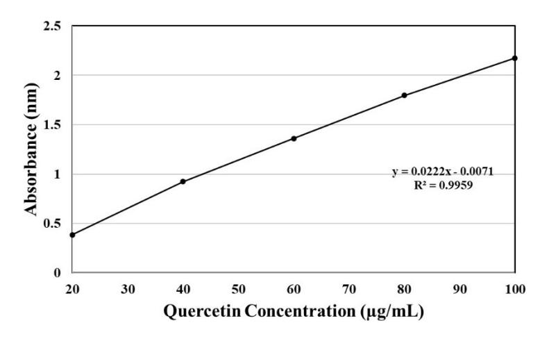
Figure 1. Quercetin Calibration Curve Acquired by Spectrophotometry
3.3 Determination of Quercetin Levels in Extract by High-Performance Liquid Chromatography
A calibration curve was made by plotting the areas under curve (AUC), as measured by the HPLC unit, of 10, 20, 40, 60, 80, and 100 μg/mL quercetin standard solutions in a linear regression with x values being a substitute for solution concentration and y values as the corresponding AUC. The equation of the calibration curve is y = 15457x – 9819.8 with a coefficient of determination value of R2 = 0.9992 (Figure 2). Using the equation of the curve, the average quercetin concentration was 11.89 µg/mL of quercetin out of 50 µg/mL solution (or 23.78% w/w), which yields 3.99 mg of quercetin per g of dried shallot skin.
3.4 Comparison of Determination Methods
From 90 g of dried shallot skin samples, 1.52 g (or 1.68% w/w) of extracts could be produced, which yielded 16.8 mg of extract per g of dried shallot skin. Determination of quercetin
content through spectrophotometry showed a yield of 3.37 mg of quercetin per g of dried shallot skin, while HPLC analysis showed a yield of 3.99 mg of quercetin per g of dried shallot skin. The discrepancy between yields resulted from differences in the analytical accuracy of the instruments used. Highperformance liquid chromatographs have a higher resolution and could differentiate different compounds contained within a sample through chromatography [36].
The results of quercetin content determination bore a resemblance with previous research [39] that shows a quercetin content of 3.42 mg per g of shallot skin; the extraction was done conventionally by centrifugation. Shallot skin would yield more flavonoids than other parts of the plant; the quercetin yield may vary from 1 to 46 mg per g of shallot skin [40, 41]. However, differences in quercetin yield could be caused by differences in plant variety, extraction method, and analysis instrument. Prior research stated that each shallot variety contained different levels of quercetin [42]. This was
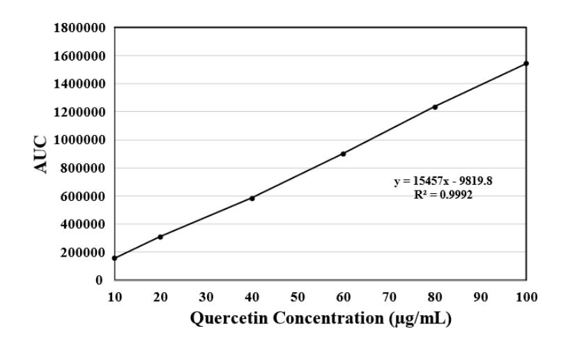
Figure 2. Quercetin Calibration Curve Acquired by HPLC
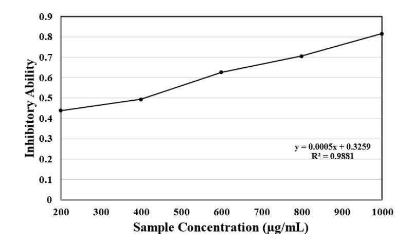
Figure 3. α-Amylase inhibition percentage by shallot skin extract
shown in their research where Allium cepa L var. Ambition had a quercetin content of 5.236 + 0.428 mg/g while Allium cepa L. var. Matador had a quercetin content of 2.258 + 0.258 mg/g. Shallots had a quercetin content of 66.33 mg/g, a higher yield compared to chartreuse onions that contained 63.71 mg/g and onions that contained 16.71 mg/g [43]. Extracting flavonoids using the subcritical water method on the Horcal variety could give maximum results at a temperature of 145°C [44]. Subcritical water extraction could yield 27.4 + 0.9 mg of quercetin per g of dried shallot skin, a higher value compared to conventional 70% ethanol extraction that only yielded 20.4 + 0.2 mg of quercetin per g of dried shallot skin.
3.5 Quercetin and Acarbose α-Amylase Inhibitory Properties Test Results
Testing was conducted by measuring the absorbance of α-amylase inhibition by shallot skin extract sample, acarbose sample, shallot skin extracts blank solution, acarbose blank solution, and control solution. Sample absorbance measured the ability of inhibitor compounds in inhibiting α-amylase
enzymatic reactions. Blank absorbance measured absorbance errors that was caused by the presence of dissolved solids while overlooking the enzymatic reaction itself. Control absorbance measured the full enzymatic activity of α-amylase without any hindrance from inhibitors (i.e, shallot skin extract, acarbose) [30].
Absorbance readings by prolonged incubation were processed into inhibition percentages. The resulting end-point inhibitory properties of acarbose and shallot skin extract are shown in the figures below. The linear regression curve of the end-point α-amylase inhibitory activity of shallot skin extract has an equation of y = 0.0005x + 0.3259 with a coefficient of determination value of R 2 = 0.9881 (Figure 3), while the curve for acarbose has an equation of y = 0.0005x + 0.3813 with a coefficient of determination value of R2 = 0.9915 (Figure 4).
The equations of the linear regressions were used to calculate the end-point IC50 value of shallot skin extract and acarbose in inhibiting α-amylase. The smaller the IC50 value of a compound is, the more potent said compound is in inhibiting a certain biochemical reaction. The end-point IC50 value of
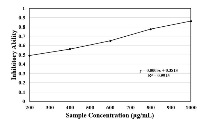
Figure 4. α-Amylase inhibition percentage by acarbose
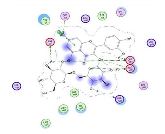
Figure 4. Molecular mechanism of α-amylase inhibition by quercetin [49] (Bamigboye et al., 2023)
shallot skin extract in inhibiting α-amylase is 348.2 μg/mL, while acarbose has an end-point IC50 value of 237.4 μg/mL. From the tests, acarbose was a more effective drug compared to shallot skin extract in inhibiting α-amylase. Pure quercetin could inhibit α-amylase with an IC50 value of 57.37 μg/mL [7]. Allium cepa L. skin extract could inhibit α-amylase with an IC50 value of 455.6 μg/mL [33]. The extract that was synthesized in this research has a lower IC50 compared to previous research [33] due to a difference in extraction method. For the same type of solvent (i.e. 70% ethanol), different durations and instruments of extraction were used. In this research, extraction was conducted by macerating the raw materials for 24 hours, followed by evaporation using rotary vacuum evaporator, while previous research [33] used thermo-shaker and ultrasonic bath with varying durations up to 2 hours and 30 minutes respectively. This caused differences in quercetin levels. Our extraction yielded higher quercetin content (3.99 mg/g) compared to previous research (3.80 mg/g). Further research is needed to devise a new extraction method in order to obtain higher quercetin yield.
3.6 Inhibitory Mechanism
α-Amylase inhibitors are compounds that prevents α-amylase from hydrolysing starch into simple sugars. In the medical field, α-amylase inhibition is done to prevent hyperglycaemia [45]. Quercetin is a flavonoid compound that can be used as an alternate α-amylase inhibitor. According to previous research using enzyme kinetic analysis, multispectroscopy, and molecular docking analysis, quercetin inhibits α-amylase competitively by forming hydrogen bonds with the active sites of the enzyme. α-Amylase active sites are located at the Glu233, Asp197, and Asp300 amino acids; by bonding with these amino acids, quercetin would prevent α-amylase from bonding with its substrate (i.e. starch) (Figure 5) [46].
3.7 Potential of Quercetin in Type 2 Diabetes Mellitus Treatment
The ability of quercetin to inhibit α-amylase makes it possible to be used as a therapeutic drug in treating type 2 diabetes mellitus (T2DM). T2DM may happen when cells that constitute muscular and organ tissues become resistant to insulin. T2DM can also happen when the human pancreas could not produce enough insulin to control blood sugar levels; this leads to the decrease of sugar uptake in the human body [48]. Glucose is made by hydrolysing starch with α-amylase as the catalyst. In the human body, α-amylase is produced secreted to the small intestines by the pancreas. As a T2DM therapeutic drug, quercetin is used to inhibit starch hydrolysis by α-amylase in the small intestines [49]. Oral administration of 500 mg or more quercetin daily for at least eight weeks can reduce fasting blood sugar levels in the human body [48]. Besides inhibiting α-amylase, as a T2DM therapeutic drug, quercetin could also induce insulin production by the pancreas, activate adenosine monophosphate kinase (AMPK) enzyme that increases cellular glucose uptake, as well as preventing insulin resistance in body tissues [50, 13].
4. Conclusion
Shallot (Allium cepa L.) skin contains a high level of quercetin that may be used as a raw material for quercetin extraction. This could diversify the utilization of shallot skin wastes as well as providing an alternative in T2DM treatment. From a 90 g sample of shallot, 1.52 g of extract (or 16.8 mg of extract per g of dried shallot skin) could be extracted. Spectrophotometry analysis indicates a quercetin yield of 3.37 mg/g of dried shallot skin, while HPLC analysis showed a yield of 3.99 mg/g of dried shallot skin. Shallot skin extract could inhibit α-amylase with an IC50 value of 348.2 μg/mL, while acarbose has an IC50 value of 237.4 μg/mL. In this research, compared to the obtained shallot skin extract, acarbose is more effective in inhibiting α-amylase. This lower
effectiveness was due to the relatively low quercetin yield obtained using a simple extraction method. Quercetin inhibits α-amylase competitively by forming hydrogen bonds with the active sites of the enzyme. The target active sites are located at Asp197, Glu233, and Asp300 amino acids. Further research is needed to devise a more effective extraction method in order to increase the quercetin yield of the extract, testing the effectiveness of quercetin as a T2DM therapeutic drug in vivo, and standardizing the physical and chemical properties of the extract up to medical criteria.
Acknowledgements
Authors express gratitude to The Lord Almighty in finishing this research. This work was financially supported by the Ministry of Education, Culture, Research and Technology of Republic of Indonesia and the Bandung Institute of Technology. Authors declare financial conflict of interest. Authors appreciate the assistance of Dr. Eri Mustari as research supervisor and Ela Kamelia Saleh as the technician of Laboratorium Isolasi dan Analisis Bahan Alam.