
1. Introduction
Bananas, particularly Kepok bananas (Musa balbisiana), are among the most economically significant horticultural commodities. In the Manokwari region, Kepok bananas, both fresh and processed, serve as a vital alternative staple food, complementing rice and contributing to food security. They are commonly prepared as boiled bananas, often served with fish and vegetables as a carbohydrate source to replace rice. Additionally, Kepok bananas are widely processed into various products, including fried bananas and banana chips, further enhancing their versatility and economic value.
According to [1] the development of banana processing businesses offers numerous benefits, including enhancing the added value of bananas compared to their fresh form, increasing farmers' income, extending storage life, and reducing postharvest losses and damage. Additionally, processing bananas
supports the efficient utilization of harvests, contributes to food diversification, and generates profitability, enabling competitiveness in both domestic and international markets. These advantages underscore the importance of ensuring the sustainable availability of bananas.
Kepok banana cultivation is highly susceptible to disease. According to data from the Agriculture Service of Sikka Regency, Flores Island, East Nusa Tenggara (NTT), as of April 2024, blood disease or bacterial wilt has affected Kepok bananas across 10 sub-districts. The infected area spans 25.62 hectares, accounting for 1.9% of the total cultivated land area of 1,348.63 hectares [2]. Blood disease is caused by the bacterium Ralstonia solanacearum race 2, with symptoms including the yellowing and wilting of one to three leaves, stem breakage, and fruit that remains green but exhibits
internal rot and mucilage upon cross-sectioning. This disease has significantly reduced banana production, contributing to NTT's inflation in March 2024 [3].
Banana diseases began to spread rapidly in Manokwari in 2018, particularly in the SP IV Bowi Subur area of Masni District [4]. Banana farmers in the Masni District are facing significant challenges due to the outbreak of banana blood disease, caused by the Blood Disease Bacterium (Ralstonia syzygii subsp. celebesensis). This disease has severely impacted banana cultivation, with damage exceeding 70% in Kepok banana varieties [5]. Consequently, the outbreak has led to a substantial decline in banana production, posing a serious threat to local livelihoods and food security.
Fusarium wilt, caused by the fungus Fusarium oxysporum f. sp. cubense, has also been reported in Masni District, resulting in losses exceeding 35% of total banana production [6]. This soil-borne disease primarily targets the roots and stems of banana plants and is notoriously difficult to manage. Symptoms of Fusarium wilt include yellowing of leaves, stem cracking, discoloration of the vascular tissues, and a foul odor emanating from infected stems [7].
In 2019, banana diseases were first reported in the Anday area of South Manokwari District. Affected plants exhibit symptoms such as rapid wilting and yellowing of leaves, with the bases of the leaves breaking. Additionally, the young fruit turns blackish-brown when cut, leading to a significant reduction in productivity [8]. The aim of this study is to isolate and identify the pathogenic bacteria responsible for Blood diseases in Kepok bananas from Anday, South Manokwari, based on their morphological and biochemical characteristics.
2. Methodology
2.1 Data collection
Samples of pseudostems, rhizosphere soil, and diseaseinfected banana fruits were collected from Anday, South Manokwari District. Pathogenic bacteria responsible for banana diseases were tested in December 2024 at the Microbiology Laboratory of FMIPA, Papua University.
2.2 Isolation of Bacteria from Pseudostems and Fruits
Bacteria were isolated from diseased banana tissues, specifically from ooze and red mucus present on pseudostems and fruit bunches. The ooze on the surfaces of fruit bunches and pseudostems was sterilized by spraying with 70% ethanol. The tissues were then longitudinally sectioned using a sterile knife into approximately 2 cm² pieces, further chopped into smaller fragments, and placed in a 2 mL tube containing 1 mL of sterile water. The samples were incubated for 5–10 minutes. Using the spread plate method, approximately 40 μL of the resulting inoculum was inoculated onto Casamino Peptone Glucose Agar (CPG) medium and incubated at room temperature for 72 hours [9].
2.3 Isolation of Bacteria from Rhizosphere Soil Samples
Composite soil samples were collected from the rhizosphere zone at a depth of 5 cm. A 10 g subsample was mixed with 90 mL of 0.9% physiological saline solution, vortexed to create a 10⁻¹ dilution, and subjected to serial dilutions up to 10⁻³. From each dilution, 1 mL was inoculated onto petri dishes containing CPG medium using the spread plate method. The plates were incubated at room temperature for 72 hours. Bacterial colonies were characterized based on color, shape, size, surface texture, elevation, and margin properties. Purification was performed on CPG medium to obtain single colonies, which were then used to prepare stock cultures and 24-hour working cultures. These cultures were subjected to Gram staining and biochemical tests, including Nutrient Agar (NA), Triple Sugar Iron Agar (TSIA), Sulfide Indole Motility (SIM), Methyl Red-Voges Proskauer (MR-VP), Simmons Citrate Agar (SCA), and Nutrient Gelatin (NG) assays, following the protocol of [10]. Nitrate reduction was assessed using the method described by [11].
2.4 Isolation of Bacteria Pathogens
Pseudostem samples were cultured on Sucrose Peptone Agar (SPA) medium and incubated for 48 hours, resulting in bacterial colonies with convex, mucoid, and milkywhite characteristics (Figure 3). The pathogenic bacteria were further purified on CPG medium supplemented with Triphenyl Tetrazolium Chloride (TTC), where the bacterial colonies developed a pink to maroon coloration (Figure 4). These findings are consistent with the results reported by [12].
2.5 Gram Staining
The Gram staining technique was employed to determine the morphological characteristics and Gram reaction of bacterial cells. A 24-hour pure bacterial culture was smeared onto a clean glass slide and heat-fixed. The smear was then stained with crystal violet for 60 seconds, followed by rinsing with distilled water to remove excess dye. Ammonium oxalate was applied to the smear for 60 seconds, rinsed off with distilled water, and the slide was decolorized by applying 96% alcohol for 10 seconds. After rinsing again with distilled water, the smear was counterstained with safranin for 30 seconds. Excess dye was removed with a final rinse of distilled water, and the slide was air-dried. Microscopic observations were conducted under 1000x magnification using immersion oil. The staining results distinguished bacterial types: Gram-positive bacteria appeared purple, while Gram-negative bacteria appeared pink [10].
2.6 TSIA Testing
Bacterial isolates were inoculated onto Triple Sugar Iron Agar (TSIA) medium (65 g/L) by stabbing the medium vertically into the butt and streaking the surface of the slant in a zig-zag pattern. The cultures were then incubated at room temperature for 48 hours, and color changes in the medium were observed. A red slant with a yellow butt indicated that the bacteria were capable of fermenting glucose only. Conversely, a yellow slant and butt indicated the bacteria's ability to ferment glucose, lactose, and sucrose [10].
2.7 Indole and Motility Test
Bacterial isolates were inoculated into Sulfide Indole Motility (SIM) medium by stabbing vertically with a straight loop and incubated at room temperature for 48 hours. The indole test was conducted to evaluate the ability of bacteria to produce the enzyme tryptophanase, which catalyzes the oxidation of the amino acid tryptophan to form indole. After incubation at 37°C for 48 hours, Kovac's reagent was added to the medium, and the formation of a cherry-red or brick-red ring on the surface of the SIM medium indicated a positive indole test result. For the motility test, bacterial movement was assessed based on the pattern of growth following the stab inoculation. Growth restricted to the stab line indicated non-motile bacteria, whereas diffusion of growth spreading laterally from the stab line suggested motility [10].
2.8 Methyl Red-Voges Proskauer Test
A single dose of the bacterial isolate was inoculated into Methyl Red-Voges Proskauer (MR-VP) medium and incubated at room temperature for 48 hours. The MR test was conducted to detect mixed acid fermentation, specifically the production of stable acids such as methylglyoxal. In contrast, the VP test assessed the ability of bacteria to produce neutral end products, such as acetoin (acetyl methyl carbinol), from glucose metabolism. For the MR test, three drops of methyl red reagent were added to the medium, and a positive result was indicated by a color change to red, signifying the presence of acidic end products. The VP test involved adding 5% α-naphthol and 40% potassium hydroxide (KOH) to the medium, followed by shaking the tube for 30 seconds. A positive VP test was determined by the appearance of a color change, indicating the production of neutral compounds [10].
2.9 Citrate Test
Asingle bacterial cycle was streaked onto Simmon's Citrate Agar (SCA) medium and incubated at room temperature for 48 hours. The test was performed to assess the ability of the bacteria to utilize citrate as a sole carbon and energy source. Color changes in the medium were observed as an indicator of citrate utilization [10].
2.10 Gelatin Hydrolysis Test
A single cycle of bacterial isolates was inoculated into Nutrient Gelatin (NG) medium and incubated at 37°C for 48 hours. After incubation, the samples were stored at 4°C for 30 minutes. Gelatin that has been hydrolyzed remains in a liquid state even when stored at 4°C. Certain microorganisms possess the ability to hydrolyze gelatin due to the production of the proteolytic exoenzyme gelatinase [10].
2.11 Nitrate Reduction Test
Nitrate broth is utilized to assess the ability of bacteria to reduce nitrate. Bacteria that are capable of rapid nitrate reduction exhibit fast growth rates, often with a short generation time. To perform the test, 0.5-1.0 mL of nitrate broth is added to a clean test tube, which is then autoclaved for 15 minutes at 1 atm pressure and 121°C. After autoclaving, allow the broth to cool to room temperature. Inoculate the tube with a heavy inoculum of fresh bacterial culture and incubate at 35°C for 2 hours. Subsequently, add two drops of reagent A (sulfanilic acid 0.8%) and two drops of reagent B (N, N-dimethyl-α-naphthylamine 0.5%), then mix thoroughly. Finally, observe the development of a red color within two minutes, which indicates a positive result [11].
3. Results and Discussion
3.1 Diseases Affecting Banana Plants
Sampling of banana plants exhibiting symptoms of the disease was conducted at coordinates 0093121" LS – 13400124" LU in Anday, South Manokwari District, Manokwari Regency, West Papua. A randomized sampling method was employed, as illustrated in Figure 1.
The observations of diseased banana plants were characterized by the presence of red mucus on the pseudostem and a blackish discoloration of young banana fruits when cut. In addition, diseased plants exhibited blackish-brown streaks on the true stem and red mucus on the pseudostem, consistent with the findings reported by [4]. Field observations revealed that the sap from cut banana stems affected by the disease turned blackish-brown within 30 minutes of being cut (Figures 2a and 2b). In contrast, no color change was observed in the sap of healthy banana stems (Figure 2c). Additionally, crosssectional slicing of the pseudostems of diseased banana plants revealed the presence of brownish-red to blackish-red mucus (Figures 2d and 2e). Soil samples, pseudostems exhibiting red slime, and banana specimens were collected and placed in labeled plastic bags. The samples were then stored in a box containing dry ice and maintained at a temperature of 4°C. Bacterial isolation procedures were initiated for each sample within 48 hours of collection.
Composite soil samples from two locations were cultured on CPG medium and incubated for 72 hours. Pathogenic bacteria isolated from the rhizosphere of mucoid banana pseudostems at Location 1 are presented in Figure 5a, while those from the rhizosphere of non-mucoid banana pseudostems at Location 2 are shown in Figure 5b. The isolation of pathogenic bacteria from bananas on CPG medium revealed consistent bacterial characteristics, including a convex shape, maroon coloration,
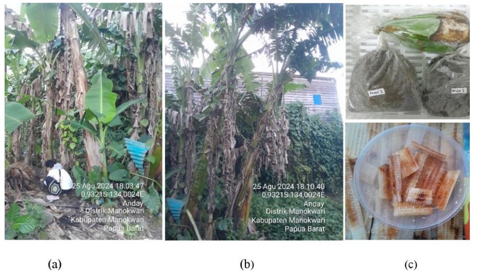
Figure 1.Rhizosphere soilsampling conducted atLocation 1 (a) andLocation 2 (b), along with the collection ofsoilsamples, banana fruits, and pseudostems(c).
and a mucoid texture. Ayesha et al reported that Blood Disease Bacteria (BDB) isolated from bananas infected with blood disease and cultured on TTC medium exhibited a reddish coloration and a mucoid texture [8]. The pathogenic bacterial isolates were assigned specific codes based on their origin (Table 1). Subsequently, stock cultures and working cultures were prepared for Gram staining and biochemical test.
3.2 The morphology of the isolates
The morphological characteristics of the isolated bacteria are summarized in Table 2. The macroscopic examination revealed that all four isolates exhibited a circular shape with smooth edges, a convex surface, and a maroon pigmentation when cultured on CPG medium. These findings align with the observations reported by [8], who described similar isolates as having a round shape, a convex surface, a red center within the colony, distinct edges, and an adhesive tendency to the surface. Isolate AR1 was obtained from the rhizosphere of a banana plant with a reddish-brown, slimy pseudostem at location 1 (Figure 1a). Isolate APS1 was collected from the pseudostem of a banana plant infected with reddish-brown slime at the same location (Figure 1a). Isolate AR2 originated from the rhizosphere of a banana plant with a healthy pseudostem at location 2 (Figure 1b). Finally, isolate ABF2 was obtained
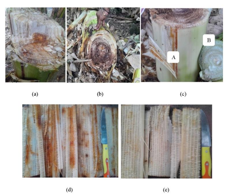
Figure 2. Vascular tissue of a blackish-brown banana stem (a), brownish-red liquid observed in the pith of the pseudostem (b), comparison between the stem of a diseased Kepok banana (A) and a healthy Kepok banana (B) (c), transverse section of the pseudostem of a diseased Kepok banana (d), and transverse section ofthe pseudostemof ahealthyKepok banana (e).
| Table 1. Designation of isolate codes based on their origins |
|---|
| No. | Isolate codes | Isolate origin |
|---|---|---|
| 1 | AR1 | Rhizosphere of banana plants with reddish-brown slimy pseudostems at Location 1 (Figure 1a) |
| 2 | APS1 | Pseudostem of a banana plant infected with reddish-brown slime at Location 1 (Figure 1a). |
| 3 | AR2 | Rhizosphere of banana plants with entirely healthy stems at Location 2 (Figure 1b) |
| 4 | ABF2 | Banana fruit sampled from Location 2 (Figure 1c) |
from banana fruit at location 2 (Figure 1c).
According to [13], Blood Disease Bacteria (BDB) exhibits specific characteristics, including a size of approximately 0.5 × 1.0–1.5 µm, a rod-shaped morphology, Gram-negative staining, lack of active motility, and the presence of flagella. Its colonies grow slowly, are non-fluidic, possess flat edges, and display a dark red center on TZC medium. Furthermore, BDB colonies are non-fluorescent when cultured on King's B medium. These findings are consistent with the results of this study, where the bacteria exhibited a red coloration on TTC-containing medium (CPG medium) and a milky white coloration on non-TTC medium (SPA medium). This is illustrated in Figure 3 and 4.
The research findings revealed that the bacterial isolates exhibited microscopic characteristics typical of Gramnegative, short rod-shaped (bacil) bacteria, as determined by Gram staining. The decolorization step using ethyl alcohol (Gram C) dissolved lipids in the outer membrane of the bacterial cell wall, increasing its permeability. Consequently, the cells retained the counterstain safranin (Gram D), resulting in a distinctive pink coloration [10].
3.3 Biochemical Test for Pathogenic Bacteria
Biochemical tests for pathogenic bacteria were conducted over a 48-hour incubation period, following the protocols outlined by [10]. The isolates were tested using the assigned codes AR1, APS1, AR2, and ABF2, corresponding to test numbers 1, 2, 3, and 4, respectively.
The bacterial testing results on Triple Sugar Iron Agar (TSIA) media demonstrated that all four isolates were capable of fermenting dextrose, lactose, and sucrose, as indicated by gas production. Gas production was evidenced by the presence of bubbles and the lifting of the media. Notably, isolate AR1 exhibited hydrogen sulfide (H₂S) production, as indicated by the blackening of the media caused by the precipitation of insoluble iron sulfide.
The carbon source utilization test, conducted using citrate on Simmons' Citrate Agar (SCA) medium, revealed positive results, indicated by a color change in bromothymol blue, the pH indicator. The findings demonstrated that the isolates AR1, APS1, AR2, and ABF2 were capable of utilizing citrate as their sole carbon and energy source in the absence of glucose or lactose.
The Voges-Proskauer (VP) test was performed to assess the ability of bacterial isolates to produce neutral (nonacidic) end products as a result of glucose metabolism. The test involved the addition of α-naphthol and 40% potassium hydroxide solution, which would result in a red coloration of the medium in the presence of neutral metabolic byproducts. However, all four isolates exhibited a negative reaction, as indicated by the absence of any color change.
The detection of mixed acid fermentation, specifically the production of methylglyoxal, was assessed in the test isolates using the Methyl Red (MR) test, with glucose serving as the primary energy substrate. This process results in the formation of acidic products, leading to a reduction in the pH of the medium. Upon the addition of the methyl red reagent, a red coloration in the medium indicates a significant pH decrease due to the mixed acid products. All four isolates (AR1, APS1, AR2, and ABF2) exhibited a positive reaction, as evidenced by the medium turning red.
Indole testing was conducted to evaluate the ability of the bacterial isolates to produce the enzyme tryptophanase, which catalyzes the degradation of tryptophan to indole. This
Table 2. Morphological characteristics of csolates collected from the rhizosphere, pseudostem, and banana fruit of Kepok banana (Musa balbisiana)
| Macroscopic characteristics | Microscopic characteristics | |||||
|---|---|---|---|---|---|---|
| Isolate code | Form | Margin | Elevation | Pigmentation | Shape | Gram stained cell |
| AR1 | circular | entire | convex | maroon | rod | negative |
| APS1 | circular | entire | convex | maroon | rod | negative |
| AR2 | circular | entire | convex | maroon | rod | negative |
| ABF2 | circular | entire | convex | maroon | rod | negative |
Figure 3.Growth of pathogenic bacteria on SPAmediumafter 24 hours ofincubation (a) and 48 hours ofincubation (b).
Figure 4. Pathogenic bacteria cultured onCPGmediumfrominfected banana pseudostemsamples after 72 hours of incubation.
was achieved by adding Kovac's reagent to the SIM (Sulfide-Indole-Motility) medium, resulting in a cherry red to brick red coloration in the presence of indole. The results demonstrated that isolates AR1, APS1, AR2, and ABF2 did not exhibit any color change, indicating a lack of indole production. Additionally, the SIM medium was utilized to assess bacterial motility, and all four isolates displayed motile behavior. The formation of a black precipitate in the SIM medium signifies hydrogen sulfide (H₂S) production. Except for isolate AR2, none of the other isolates produced H₂S, as evidenced by the absence of black coloration.
Gelatin hydrolysis testing, performed using nutrient gelatin media, revealed that isolates AR1, AR2, and ABF2 were unable to hydrolyze gelatin, as the medium solidified after incubation at 4°C for 30 minutes. In contrast, the medium inoculated with isolate APS1 remained liquefied, suggesting that APS1 possesses the ability to hydrolyze gelatin.
Nitrate reduction activity was assessed using nitrate broth medium, with the addition of reagent A (0.8% sulfanilic acid) and reagent B (0.5% N,N-dimethyl-α-naphthylamine). This rapid assay detects nitrate reduction through a color change in the medium to red after the addition of two drops each of reagents A and B. The test results indicated that none of the isolates (AR1, APS1, AR2, and ABF2) exhibited nitrate reduction, as no color change was observed.
The catalase test results demonstrated that all four isolates
produced the enzyme catalase, as evidenced by the formation of air bubbles when hydrogen peroxide (H₂O₂) was applied to glass slides smeared with the bacterial isolates. The catalase activity varied among the isolates, increasing in the order: AR1, APS1, AR2, and ABF2. In conclusion, the isolates AR1, APS1, AR2, and ABF2 exhibit nearly identical characteristics. The comprehensive results of the bacterial biochemical tests are summarized in Table 3. Isolates AR1, APS1, AR2, and ABF2 share several characteristics, including the ability to ferment dextrose, lactose, and sucrose, as well as to produce gas. They are capable of utilizing citrate as a carbon and energy source in the absence of glucose or lactose, do not produce neutral (non-acid) end products, and use glucose as the primary substrate for energy, resulting in the production of acidic byproducts that lower the pH of the medium. Additionally, these isolates do not produce the enzyme tryptophanase, are motile, are unable to reduce nitrate, and can produce the enzyme catalase. Notably, isolate APS1 was the only one capable of hydrolyzing gelatin, while isolate AR2 was the only one that did not produce H₂S.
Three types of bacterial wilt disease have been reported in banana plants: Moko disease, caused by Ralstonia solanacearum race 2, endemic to Central and South America, the southern Philippines [4,14-15]. Bugtok disease, also caused by Ralstonia solanacearum race 2 in the Philippines; and Blood disease, caused by Ralstonia syzygii in Indonesia
Figure 5. Pathogenic bacteria cultured onCPGmediumafter a 72-hourincubation period:(a)isolated fromthe rhizosphere ofmucoid pseudostems, and (b)isolated fromthe rhizosphereofnon-mucoid pseudostems
[16]. In a new classification system based on molecular analysis, Blood disease, caused by Ralstonia syzygii, is classified in phylotype IV (Indonesia) according to its origin and evolutionary history [4,16-17]. In addition to Indonesia, Ralstonia solanacearum phylotype IV of this species complex has also been isolated from Australia, Japan, Korea, and Malaysia. Following taxonomic and nomenclatural revisions, this phylotype was reclassified as Ralstonia syzygii, which was further divided into three subspecies: Ralstonia syzygii subsp. syzygii, the pathogen responsible for disease in clove trees in Sumatra; Ralstonia syzygii subsp. Indonesiensis, the pathogen causing bacterial wilt disease in various host plants; and Ralstonia syzygii subsp. celebesensis, the pathogen responsible for blood disease in Musa spp. [17]. Banana disease was first identified on Selayar Island, South Sulawesi, in 1910. For decades, its spread was restricted to this region due to strict quarantine measures enforced by the Dutch [4,16]. However, the disease eventually spread across Sulawesi and was detected in Java by the 1980s. Its transmission to sparsely
populated islands was facilitated by transmigration. In regions affected by banana blood disease, control measures include the early removal of male flowers and the wrapping of fruit bunches to prevent further spread [16].
Prior et al stated that R. solanacearum, R. syzygii, and Ralstonia syzygii subsp. celebesensis exhibit distinct geographic distributions and pathogenic potentials, and can be easily differentiated genetically [18]. When considering a single denitrification metabolic pathway, members of this third group can be classified as subspecies. Although genetically related, these three taxa are distinguishable based on their geographic distribution and differing pathogenic potentials.
Endemic banana plantations become unproductive due to contamination by infective propagules of the blood disease bacterium. This represents a significant obstacle to the rehabilitation of banana plantations in Indonesia. The infective propagules of BDB can persist in soil and infected plant tissue for up to 1–2 years, retaining their virulence [1].
The characteristics of BDB differ slightly from those of R.
Table 2. Biochemical Test Results for Bacterial Isolates from Rhizosphere, Pseudo-stem, and Banana Fruit Samples
| Isolate | |||||
|---|---|---|---|---|---|
| Parameter | AR1 | APS1 | AR2 | ABF2 | |
| Fermentation of Sucrose, Lactose, and Dextrose | + | + | + | + | |
| Citrate Test | + | + | + | + | |
| Voges Proskauer test | - | - | - | - | |
| Methyl Red Test | + | + | + | + | |
| Motality | - | - | - | - | |
| H2S Production | + | + | - | + | |
| Indol Production | - | - | - | - | |
| Gelatin Hydrolysis | - | + | - | - | |
| Nitrate Reducers | - | - | - | - | |
| Catalase Test | + | ++ | +++ | ++++ | |
+ : Able to produce
- : Doesn't produce
++ : Able to produce more
+++ : Capable of producing a lot
++++ :Capable of producing a very large amount
solanacearum. While BDB is phenotypically and genetically distinct from R. solanacearum, it shares physiological similarities with it. The primary difference is that the pathogen responsible for blood disease is unable to reduce nitrate to nitrite and cannot hydrolyze gelatin [13].
Biochemical test results indicated that isolates from the rhizosphere of Location 1 and Location 2 (AR1, AR2), as well as the isolate from banana fruit at Location 2 (ABF2), were all unable to reduce nitrate and hydrolyze gelatin. Therefore, these isolates can be identified as blood disease bacteria (BDB), specifically Ralstonia syzygii phylotype IV (Ralstonia syzygii subsp. celebesensis).
The isolate derived from the pseudostem at Location 1 (APS1) was unable to reduce nitrate but exhibited the ability to hydrolyze gelatin. This characteristic distinguishes APS1 from isolates AR1, AR2, and ABF2, which cannot hydrolyze gelatin. Therefore, APS1 is likely not a blood disease bacterium (Ralstonia syzygii subsp. celebesensis). However, due to its inability to reduce nitrate to nitrite, APS1 cannot be classified as R. solanacearum.
4. Conclusion
The isolation of samples from the banana rhizosphere, pseudostem, and banana fruit yielded four isolates: AR1, APS1, AR2, and ABF2. Based on nitrate reduction and gelatin hydrolysis tests, isolates AR1, AR2, and ABF2 were identified as blood disease bacteria (Ralstonia syzygii subsp. celebesensis). In contrast, the APS1 isolate could not be classified as either Ralstonia syzygii subsp. celebesensis or R. solanacearum.
Acknowledgements
We sincerely appreciate the Master of Biology Program at PPs Unipa for providing access to laboratory equipment, which was essential for the successful completion of this research.