

1. Introduction
Shrimp is a leading commodity in aquaculture due to its high economic value in both export and local markets [1]. Despite the potential, several shrimp species that are economically important and widely cultured in Indonesia are often affected by Vibrio infections. This has led to a decrease in production, particularly for tiger shrimp (Penaeus monodon) [2]. The decline in tiger shrimp production is due to disease attacks caused by bacteria at the larval stage. Poor physical and chemical parameters often cause bacterial infection and increase mortality in shrimp [3].
Vibrio harveyi is a pathogenic bacteria that frequently infects shrimp and fish larvae, causing various diseases, such as luminescent vibrio or vibriosis[4]. A common strategy to control V. harveyi infections is antibiotic administration, but long-term use can lead to antibiotic resistance [5]. Therefore, natural compounds with antimicrobial properties are being explored as alternative treatments.
Several studies on antimicrobial application assay of natural ingredients have been conducted, such as cloves [6], cinnamon [7], pepper [8], citrus extracts [9], garlic [10], and K. alvarezii [11].
K. alvarezii is a type of seaweed that has been widely used as a source of food, medicine, and cosmetic ingredients [12]. It has also been reported to contain secondary metabolite compounds that can produce antibacterial activity [13], such as phenol and its derivatives (flavonoid). These compounds damage the cytoplasmic membrane with H+ ions, leading to inhibited growth and death [14].
Several studies have used K. alvarezii extracts to inhibit bacterial growth, potentially leading to cell death in both gram-negative and gram-positive bacteria. In addition, the bioactivity of the extract tends to be immunomodulatory [15]. Therefore, this study aims to characterize various types of natural ingredients that have the potential to be used as antibacterial in shrimp ponds. The use of natural ingredients is one of the breakthroughs to gradually replace the use of chemical-based antibiotics that are widely used.
2. Methodology
The tools used in this study included blender, tray, analytical balance, spatula, macerator, beaker glass, evaporator, oven, petri dish, desiccator, ose needle, test tube, tweezers, incubator, measuring cup, micro pipette, tube rack, lighter, hot plate, stirrer bar, autoclave, drop pipette, Erlenmeyer flask, porcelain cup, funnel, vortex and Benson burner. The materials used were K. alvarezii, TCBS (Thiosulfate Citrate Bile Salts Sucrose Agar), distilled water, disc paper, filter paper, aluminum foil, 2N sulfuric acid, iodine, Kl, methanol, sulfuric acid, acetic acid, 5% FeCl3, 2N HCl, magnesium powder, amyl alcohol, 70% alcohol, 1% FeCl, peptone, NaCl and V. harveyi.
Seaweed seed used in this study was 45 days old, obtained from traditional farmers and under the supervision of Laboratorium Mini Hatchery, Faculty of Fisheries and Marine Science, Universitas Borneo Tarakan. Furthermore, seaweed extract was obtained from healthy thallus tissue, characterized by a complete structure, absence of slime or white spots, opacity, and resistance to breakage. V. harveyi was obtained from the Balai Besar Perikanan Budidaya Air Payau (BBPBAP) Jepara.
2.1 Seaweed Extract (Kappaphycus alvarezii)
K. alvarezii extract (Figure 1) was processed using the maceration or soaking method [16], washed and dried for 5-7 days, then pureed. Homogenized seaweed was macerated using 70% ethanol for 3 days at room temperature. The filtrate was obtained by filtration and evaporated using rotary evaporator to obtain a concentrated extract.
2.2 Phytochemical Assay
2.2.1 Alkaloid Assay
Alkaloid assay was carried out using Wagner's reagent, which consisted of 2.5 g iodine and 2 g Kl, dissolvedin 100 ml of distilled water. For the test, 0.01 g of extract was dissolved in a few drops of 2N sulfuric acid, followed by addition of two drops of Wegner's reagent. The presence of alkaloids was indicated by the formation of a brown precipitate [17].
2.2.2 Phenol Assay/FeCl3 Solution
For the phenol assay, 0.01 g of extract was dissolved in 1 ml of 70% ethanol, and two drops of 5% FeCl3 were added. Positive results were characterized by the formation of green or blue green color [17].
2.2.3 Flavonoid Assay
Flavonoid assay was made by dissolving 0.01 g of K. alvarezii extract and 0.1 mg of magnesium powder with 0.4 ml of alcohol (37% hydrochloric acid and 96% ethanol with the same volume). A total of 4 ml of 96% ethanol was added and the mixture was then shaken. Positive assay results were characterized by the formation of red, yellow, or orange color [17].
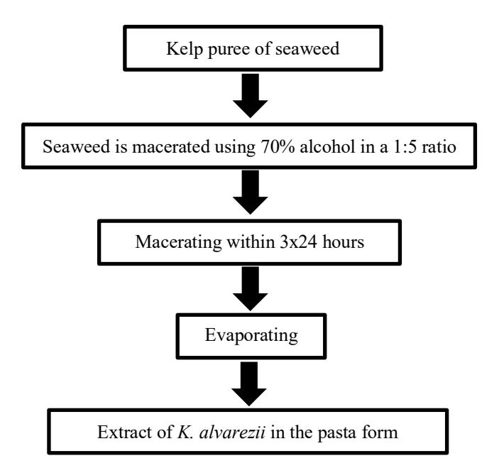
Figure1.Schematic diagramoftheK. alvarezii extraction process
2.2.4 Saponin Assay
The K. Alvarezii was dissolved in water (heating can be added to aid solubility). The solution was shaken vigorously. The formation of stable foam, persisting for several minutes and reaching a height of 1–10 cm, indicated the presence of saponins. To confirm this result, one drop of 2N HCl was added to the foamed solution; the persistence of foam further supported the presence of saponins [17].
2.2.5 Steroid Assay
Steroid assay procedure was conducted by adding 2 mg of extract to 20 ml of methanol containing 2 ml of sulfuric acid, then the mixture was boiled, followed by adding 2 ml of acetic acid. Positive results appeared when there was a change in color to green or blue [17].
2.2.6 Tannin Assay
Tannin assay procedure was conducted by dissolving the extract in distilled water and then boiled for 5 minutes. After cooling to room temperature, 3-5 drops of FeCl3 reagent 1% were added. Positive results were indicated when there was a change in color to black, blue, or green-blue [17].
2.2.7 Regeneration of Vibrio harveyi
V. harveyi originated from Balai Besar Perikanan Budidaya Air Payau (BBPBAP) Jepara. V. harveyi was inoculated into TCBS media (Thiosulfate Citrate Bile Salts Sucrose Agar) and incubated for 24 hours. Furthermore, fresh bacteria from TCBS media were inoculated into TCBS media.
Thiosulfate Citrate Bile Salts Sucrose Agar (TCBS agar) is a selective medium used to isolate Vibrio species. To prepare the medium, 89–90 grams of TCBS agar powder was dissolved in one liter of distilled water and gently heated with stirring until completely dissolved. The medium should not be instead, it only needs to be brought to a boil to sterilize. After cooling to about 45–50°C, the medium was poured into sterile Petri dishes, and allowed it to solidify. The prepared plates are stored at 2–8°C and kept protected from light. Once the agar media was solidified, the bacteria were cultured in TCBS plates and incubated at 37°C for 24 hours [18].
2.2.8 Anti-Bacterial Assay
Anti-bacterial activity assay was carried out using the scratch method on the entire surface of TCBS media and attached paper disks that had been previously soaked for 24 hours with each extract treatment.
The concentrations in this study included 5% (0.5 g), 10% (1 g), 15% (1.5 g), and 20% (2 g) while tetracycline served as the positive control and a solvent-only disc as the negative control, with 3 repetitions. Fresh cultures of V. harveyi from TCBS agar were subcultured by the pour plate technique on new TCBS plates and incubated at 37°C for 24 hours [14]. Total test tube that will use in this study as much as 15 tubes. Microbial growth and measurement of the diameter of inhibition zone were observed, characterized by the formation of a clear area around the disc paper using a caliper ruler or sigma. The diameter of inhibition zone formed on each disc paper was measured from 2 different sides. The measurements of each side were summed and divided by vertical and horizontal, then averaged, as illustrated in Figure 2 [19].
2.2.9 Data Analysis
The results of inhibition zone from the treatment were statistically analyzed using one-way ANOVA (analysis of variance) by SPSS version 23.0. Further tests were carried out using the Duncan test to determine the significance of the difference between treatment and control in means with a confidence level of 95% (margin of error 5%).
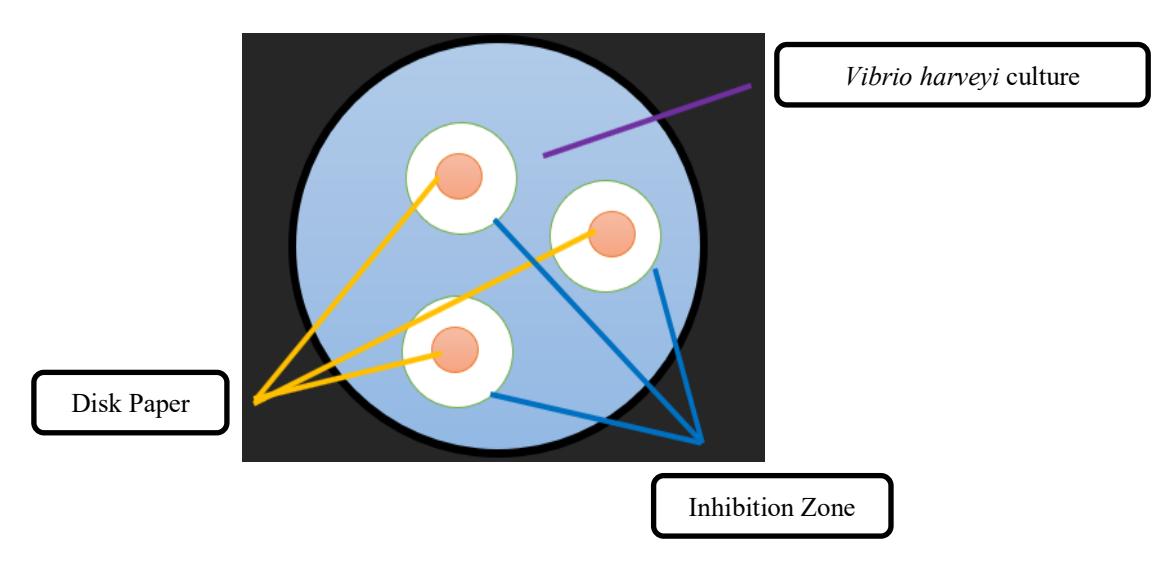
Figure 2.Inhibition zone models
3. Result and Discussion
Phytochemical compounds were secondary metabolite groups in plants that had certain functions in humans, animals, or plants [20]. The results of phytochemical analysis on K. alvarezii extract were presented in Table 1. Furthermore, it had flavonoid, alkaloid, saponin, phenol, tannin, and steroid [21]. Based on the results obtained, K. alvarezii extract had the ability as antibacterial properties. The presence of secondary metabolite compounds in the extract could disrupt the bacterial cell membrane, ensuring that V. harveyi bacteria were not able to replicate and grow [22].
Alkaloid
Phytochemical assay on K. alvarezii extract using Wegner's reagent showed a brown color, which suggested indicated the presence of alkaloid compounds. This finding is consistent with previous literature [23]. A similar result using Dragendorf reagent showed the orange formation, confirming that K. alvarezii extract contained alkaloid compounds [24]. Alkaloid compounds had the ability as antibacterial properties. The mechanism of action involves damaging bacterial cell structures and disrupting essential metabolic processes. Alkaloids can also induce DNA damage within bacterial nucleus, leading to nuclear lysis and ultimately causing cell death [25].
Phenol
Phenol referred to a functional component commonly found in flavonoid. The presence of phenol content in the extract was indicated by the formation of green color after using FeCl3 solution. Furthermore, this result is in line with previous research showing the presence of phenol content in Padina australis and Eucheuma cottoni extracts by the formation of green or blue-green color [26]. Phenol compounds (carbolic acid) act as an antibacterials by disrupting and damaging cell membrane. The acidic properties of phenol compounds could affect the growth of Streptococcus mutans bacteria [27]. Phenol compounds also could denature proteins and damage membrane integrity, thereby affecting bacterial growth [28].
Flavonoid
Phytochemical assay of K. alvarezii extract using magnesium reagent showed that K. alvarezii contained flavonoid compounds characterized by red or greenish yellow [29]. However, another study showed that K. alvarezii extract did not contain flavonoid compounds [30]. One factor that may explain these contradictory findings is the difference in extraction methods, particularly the type of solvent used. The absence of secondary metabolite compounds obtained from K. alvarezii extracts was due to the drying process of seaweed using sunlight. Furthermore, the sunlight suggested that the secondary metabolites were damaged. This was supported by the other results, showing that the phytochemical compounds of seaweed dried using sunlight and extracted using ethanol solvents, had triterpenoid compounds but did not have alkaloid compounds, flavonoid, or steroid [31].
Flavonoid are lipophilic compund capable of disrupting microbial membranes [28]. In K. alvarezii extract, the presence of flavonoid potentially act as antibacterial agent with membrane-damaging mechanism In addition,to ability in inhibiting bacterial growth, flavonoids have been reported as antimicrobial and antiviral properties [32], as well as broader bioactivites includinganti-inflammatory, antiallergic, anticarcinogenic, antioxidant, and vasoprotective effect [33]. They are widely distributed in various plant parts, such as leaves, seeds, peels, and flowers [30].
Table1.Results ofPhytochemicalAssay ofSeaweedExtract(K. alvarezii)
| No | Phytochemical Assay | Solution | Result | Description |
|---|---|---|---|---|
| 1 | Alkaloid | Wegner | + | Brown colored solution |
| 2 | Phenol | FeCl3 5% | + | Green colored solution |
| 3 | Flavonoid | HCL + Mg | + | Yellow colored solution |
| 4 | Saponin | HCl | + | Bubble formed |
| 5 | Steroid | Acetic Acid | + | Green colored solution |
| 6 | Tannin | FeCl3 1% | + | Green colored solution |
Saponin
Based on the results of phytochemical assay of saponin compounds using HCl reagent, it was found that K. alvarezii extract had saponin. Saponin was characterized by the formation of bubbles when shaken and could last for 10 minutes. Another study showed that K. alvarezii contained saponin compounds using the reagent of distilled water, which was characterized by the presence of foam [23]. Saponin had amphiphilic properties, which caused foaming because the compound could form lipid bonds with cell membranes [30].
Steroid
The results showed that the presence of steroid compounds in K. alvarezii extract was indicated by green color. This result is in line with previous research [34]. The mechanism of action of steroid compounds is by damaging the plasma membrane of bacterial cells by damaging the cell membrane, resulting in cell leakage which will cause death. Steroid had potential as antibacterial compounds with the mechanism of inhibiting protein synthesis [35], and acted as antibacterial and antifungal, which could damage bacterial cell membranes and inhibit bacterial growth. This compound, as a hormone, involved binding to specific intracellular receptors, which then regulated gene expression.
Tannin
Phytochemical assay of K. alvarezi resulted in seaweed extract containing tannin compounds characterized by the formation of green color. Another study similarly reported the presence of tannin compounds in K. alvarezii extract by the formation of blackish green color [23].
Tannin compounds functioned as antibacterials by damaging and rupturing bacterial cell walls, leading to cell death. Macroalgae extracts had alkaloid, tannin, and triterpenoid compounds that exhibited antibacterial activity
[36]. Moreover, tannin compounds are active secondary metabolite compounds that are useful as antidiarrheal, antibacterial, and antioxidants [36].
Antibacterial Test of Seaweed Extract (Kappaphycus alvarezii) against Vibrio harveyi
K. alvarezii extract has been reported to exhibit antibacterial activity against Aeromonas hydrophila and V. harveyi which was attributed to its secondary metabolite compound [37]. In this study, we evaluated this antibacterial potential against V. harveyi by measuring inhibition zone after 24 hours of incubation of different extract concentration in TBCS agar. The measurement of inhibition zone of seaweed extract was shown in Figure 3 and 4.
The largest inhibition zone was observed in the tetracycline with an average value of 33.6 mm. In comparison, the treatment with a concentration of 20%, 15%, 10%, and 5% extract was 13.3 mm, 7.3 mm, 7.1 mm, and 7.1 mm, respectively. with higher extract concentrations producing larger inhibition zones. However, tetracycline still exhibited the strongest antibacterial activity among all treatments..
Antibacterial activity with a diameter > 20 mm was categorized as strong inhibitor, diameter 11 to 20 mm was categorized as moderate inhibitor, while diameter < 10 mm was categorized as weak inhibitor [38]. Based on this classification, 5% to 15% extract concentrations were categorized as weak inhibitors, while 20% extract demonstrated moderate antibacterial activity. There are 4 factors could affect the results of antibacterial assay, namely extract concentration, secondary metabolite compound, extract diffusion power, and the type of bacteria being tested [39].
Based on ANOVA, a significant value of 0.00 <0.05 was obtained, indicating a significant difference between each treatment. In further assay, the Duncan assay showed that the 5%, 10% and 15% treatments had no significant difference,
Figure 3. Inhibition zones produced by K. alvarezii extract at different concentrations. (A) Positive control; (B) negative control; (C) 5% extract; (D) 10% extract; (E) 15% extract; and (F) 20% extract.
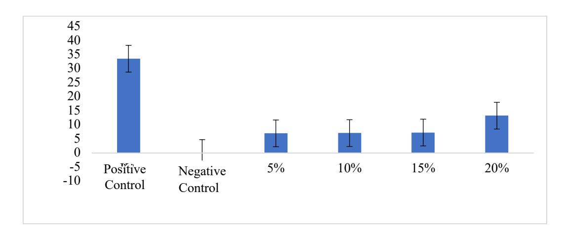
Figure4.InhibitionzoneofK. alvarezii extract againstV. harveyi
and the 20% treatment showed the best treatment against V. harveyi. In this case, higher extract concentration produced larger inhibition zone, indicating stronger antibacterial activity As the inhibition zone increased with concentration, the ability of extract to suppress bacterial growth also increased.
4. Conclusion
In conclusion, K. alvarezii extract has antibacterial ability in inhibiting V. harveyi bacteria, characterized by the presence of inhibition zone. Antibacterial ability of K. alvarezii extract largely due to the presence of phytochemical contents in the form of alkaloid, saponin, tannin, phenol, flavonoid, and steroid. Antibacterial activity of seaweed extract obtained the largest inhibition zone at a concentration of 20% with inhibition zone diameter of 13.3 mm, 15% by 7.3 mm, 10% by 7.1 mm, and 5% by 7 mm.
Acknowledgements
The author would like to thank for the Institute for Research and Community Service, University of Borneo Tarakan for the grant through DIPA 2022.